Home > Arts > Artists > P > those present
Duodenal microvilli

![]()
Wall Art and Photo Gifts from Science Photo Library
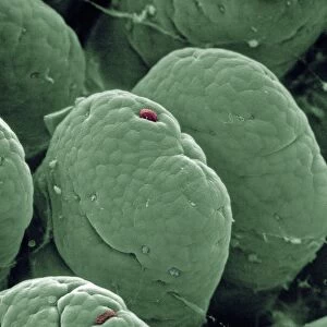
Duodenal microvilli
Microvilli in duodenum. Coloured transmission electron micrograph (TEM) of a section through the human duodenum, showing microvilli on the surface epithelium (lining). The duodenum is the first part of the small intestine. The microvilli (pink) appear as tiny projections from the surface of the epithelial cells (at lower frame). Microvilli are present on two specialised cell types that comprise the duodenal epithelium. One type, goblet cells, secrete mucus; a second type, secretory cells, secrete digestive enzymes and an alkaline fluid. The existence of microvilli serves to maximise the duodenums surface area and hence its capacity to secrete. Magnification: unknown
Science Photo Library features Science and Medical images including photos and illustrations
Media ID 6451093
© STEVE GSCHMEISSNER/SCIENCE PHOTO LIBRARY
Alimentary Canal Coloured Tem Digestion Digestive System Digestive Tract Duodenum Magnified Image Micrograph Microscopic Photos Microvilli Small Intestine Subjects System Transmission Electron
FEATURES IN THESE COLLECTIONS
> Arts
> Artists
> P
> those present
EDITORS COMMENTS
This print from Science Photo Library showcases the intricate beauty of duodenal microvilli, which are tiny projections on the surface epithelium of the human duodenum. The duodenum, as the initial segment of the small intestine, plays a crucial role in digestion within our bodies. In this colored transmission electron micrograph (TEM), we can observe these microvilli appearing pink and emerging from the surface of epithelial cells. These remarkable structures are present on two specialized cell types found in the duodenal epithelium: goblet cells and secretory cells. Goblet cells secrete mucus while secretory cells produce digestive enzymes and an alkaline fluid. The presence of microvilli serves a vital purpose - to maximize the surface area of the duodenum, thereby enhancing its capacity for secretion. This feature is essential for efficient digestion and absorption processes within our alimentary canal. Through an unknown magnification level, this image offers us a glimpse into one aspect of our complex digestive system at a microscopic level. It highlights how science allows us to explore subjects like anatomy and physiology with incredible detail, unraveling mysteries that lie beneath our skin. This print by Science Photo Library is not intended for commercial use but rather aims to educate viewers about this fascinating aspect of human biology.
MADE IN THE UK
Safe Shipping with 30 Day Money Back Guarantee
FREE PERSONALISATION*
We are proud to offer a range of customisation features including Personalised Captions, Color Filters and Picture Zoom Tools
SECURE PAYMENTS
We happily accept a wide range of payment options so you can pay for the things you need in the way that is most convenient for you
* Options may vary by product and licensing agreement. Zoomed Pictures can be adjusted in the Basket.