Fraud Blocker Skip to main content

Mucous Collection

"Mucous: The Unsung Hero of Our Respiratory System and Beyond" Nasal Lining

Background imageMucous Collection: Nasal lining, SEM

Nasal lining, SEM
Nasal lining. Coloured scanning electron micrograph (SEM) of the olfactory epithelium that lines the nasal cavity, showing olfactory cells (red) surrounded by numerous cilia (hair-like projections)

Background imageMucous Collection: Trachea cross-section showing normal and asthmatic bronchiole

Trachea cross-section showing normal and asthmatic bronchiole
Trachea cross-section showing comparison of normal and asthmatic bronchiole

Background imageMucous Collection: Trachea cross-section with lungs showing normal and asthmatic bronchiole

Trachea cross-section with lungs showing normal and asthmatic bronchiole
Trachea cross-section with lungs, showing comparison of normal and asthmatic bronchiole

Background imageMucous Collection: Small intestine, light micrograph

Small intestine, light micrograph
Small intestine. Light micrograph of a section through the small intestine. The finger-like projections at left are the villi. These increase the surface area for the absorption of food

Background imageMucous Collection: MUC7 molecule

MUC7 molecule. Computer model showing the structure of a molecule of the protein MUC7 (mucin 7). Mucins key characteristic is their ability to form gels

Background imageMucous Collection: MUC5B molecule

MUC5B molecule. Computer model showing the structure of a molecule of the protein MUC5B (mucin 5 subtype B). Mucins key characteristic is their ability to form gels

Background imageMucous Collection: Cystic fibrosis, conceptual artwork C015 / 5022

Cystic fibrosis, conceptual artwork C015 / 5022
Cystic fibrosis, conceptual computer artwork. Molecule of DNA (deoxyribonucleic acid, blue) partly covered in mucus (yellow) surrounded by salt (NaCl) molecules

Background imageMucous Collection: Stomach lining, SEM C016 / 9068

Stomach lining, SEM C016 / 9068
Stomach lining. Coloured scanning electron micrograph (SEM) of the lining of the stomach, known as the mucosa. The stomach is a muscular sac involved in storage and digestion of food

Background imageMucous Collection: Smell receptor, TEM

Smell receptor, TEM
Smell receptor. Transmission electron micrograph (TEM) of a section through the olfactory epithelium that lines the nasal cavity, showing an olfactory cell (smell receptor)

Background imageMucous Collection: Trachea lining, SEM C015 / 9610

Trachea lining, SEM C015 / 9610
Trachea lining. Coloured scanning electron micrograph (SEM) of the lining of the trachea (wind pipe). The trachea links the larynx to the lungs

Background imageMucous Collection: Tongue, light micrograph C015 / 7105

Tongue, light micrograph C015 / 7105
Tongue. Light micrograph of a section through healthy tongue tissue. The surface of the tongue (across top) consists of stratified squamous epithelium (dark purple) with a rough keratinised surface

Background imageMucous Collection: Trachea lining, SEM C015 / 9609

Trachea lining, SEM C015 / 9609
Trachea lining, SEM

Background imageMucous Collection: Anatomy in the 18th C

Anatomy in the 18th C
a skull, teeth, the interior of a knee (below left) some marrow and a mucous gland from the humerus (below right) Date: Circa 1760

Background imageMucous Collection: Snail foot

Snail foot. Foot of a garden snail (Helix aspersa) crawling over glass, seen from below. Snails have a single foot. Movement is created by successive contractions of the muscles inside the foot

Background imageMucous Collection: Small intestine villus, SEM

Small intestine villus, SEM
Small intestine villus. Coloured scanning electron micrograph (SEM) of a freeze fracture section through a villus from the mucosal lining of the small intestine

Background imageMucous Collection: Nose mucosa, light micrograph

Nose mucosa, light micrograph
Nose mucosa. Light micrograph of a section through the nasal mucosa (the tissue that lines the airways of the nose), in the region lying over the nasal concha

Background imageMucous Collection: Salivary gland, light micrograph

Salivary gland, light micrograph
Salivary gland. Coloured light micrograph of a section through a sublingual salivary gland, which is situated directly under the tongue

Background imageMucous Collection: Runny nose

Runny nose. Artwork of a section through the ear, nose and throat of a patient with excessive mucus production due to a cold or hay fever (allergic rhinitis)

Background imageMucous Collection: Nasal polyp, light micrograph

Nasal polyp, light micrograph
Nasal polyp. Light micrograph of a section through a nasal polyp, a benign growth that arises from a mucous membrane. Magnification: x150 when printed at 10 centimetres wide

Background imageMucous Collection: Trachea mucous membrane, SEM

Trachea mucous membrane, SEM
Trachea mucous membrane. Coloured scanning electron micrograph (SEM) of a fractured mucous membrane of the trachea (wind pipe), showing the epithelium and underlying connective tissue

Background imageMucous Collection: Frog skin glands, light micrograph

Frog skin glands, light micrograph
Frog skin glands. Light micrograph of a section through the skin of a frog, showing secretory glands (round, upper centre) just below the surface (across top)

Background imageMucous Collection: Nasal mucosa, artwork

Nasal mucosa, artwork
Nasal mucosa, cross-section. Artwork of a sequence (left to right) showing mucus production in response to infection and inflammation

Background imageMucous Collection: Asthma and mucus overproduction, artwork

Asthma and mucus overproduction, artwork
Asthma and mucus overproduction. Artwork showing how the bodys immune response causes the inflammation seen in asthma. The chest of a human figure is at top right

Background imageMucous Collection: Small intestine lining, light micrograph

Small intestine lining, light micrograph
Small intestine lining. Light micrograph of a section through the finger-like projections (villi) of the duodenum, the uppermost part of the small intestine

Background imageMucous Collection: Snake ciliated lung cells and mucus, SEM

Snake ciliated lung cells and mucus, SEM
Snake ciliated lung cells and mucus. Coloured scanning electron micrograph (SEM) of ciliated cells (green) and mucus (pink) in the lung of a snake


All Professionally Made to Order for Quick Shipping

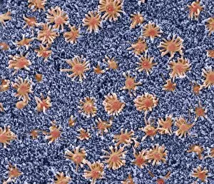
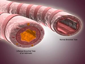
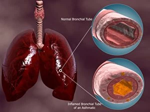
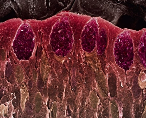
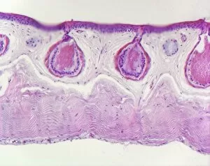
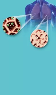
"Mucous: The Unsung Hero of Our Respiratory System and Beyond" Nasal Lining: A microscopic view reveals the intricate network of mucous-producing cells that line our nasal passages, protecting us from harmful particles. SEM Trachea Cross-Section: Explore the fascinating world within our airways as we zoom in on a cross-section, showcasing both normal and asthmatic bronchioles. Asthma's Impact: Witness the stark contrast between a healthy trachea cross-section and one affected by asthma, highlighting the crucial role mucous plays in respiratory health. Small Intestine Micrograph: Delve into the realm of digestion with a captivating light micrograph capturing the small intestine's surface covered in protective layers of mucous. MUC7 Molecule Unveiled: Meet MUC7, an essential protein found in saliva that not only lubricates but also defends against potential pathogens lurking in our mouths. Cystic Fibrosis Artwork: Step into a conceptual artwork depicting cystic fibrosis, where malfunctioning mucous production leads to severe respiratory complications – shedding light on its importance for lung health. Mighty MUC5B at Work: Discover how this remarkable molecule forms thick strands within our airways, providing additional protection against foreign invaders and maintaining their integrity. SEM Stomach Lining Exploration: Journey through an electron microscope image revealing the intricacies of stomach lining coated with mucus – shielding it from corrosive digestive acids. TEM Smell Receptor Revelation: Peer into the tiniest details as we uncover how specialized smell receptors are enveloped by mucous membranes, enhancing our olfactory experience. Invisible yet indispensable, mucous is more than just slimy secretions; it serves as nature's shield across various parts of our body - safeguarding us from harm while ensuring optimal functioning.