Framed Print > Europe > United Kingdom > Scotland > Falkirk > Bo'ness
Framed Print : Brain motor cortex pathways, artwork C016 / 6532


![]()
Framed Photos from Science Photo Library
Brain motor cortex pathways, artwork C016 / 6532
Brain motor cortex pathways. Artwork of a sectioned human brain, brainstem and spinal cord, showing neural pathways (red and blue) from the motor cortex of the brain. These are extrapyramidal pathways: the rubrospinal and reticulospinal tracts. Extrapyramidal pathways control involuntary movements and help co-ordinate movements (muscle at lower left). For an image showing the brain and where these sections were obtained, see C016/6531. The top image is a vertical (coronal) section through the brain at the rear of the frontal lobe. From top to bottom, the horizontal sections are at the level of the: mid brain, pons, medulla, and spinal cord
Science Photo Library features Science and Medical images including photos and illustrations
Media ID 9244473
© BO VEISLAND/SCIENCE PHOTO LIBRARY
Axial Axons Brain Stem Cerebral Cortex Cingulate Gyrus Coronal Medulla Medulla Oblongata Midbrain Motor Cortex Pathway Neural Pathway Parietal Lobe Physiological Physiology Pons Sections Somatosensory Cortex Spinal Cord Substantia Nigra Brain Cutouts Lentiform Nucleus Motor Cortex Neurological Neurology Section Sectioned
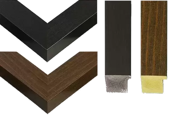
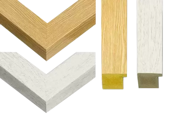
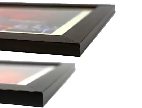
22"x18" (58x48cm) Modern Frame
Discover the intricacies of the human brain with our stunning Framed Print of "Brain motor cortex pathways" by BO VEISLAND/SCIENCE PHOTO LIBRARY. This captivating artwork showcases a detailed sectioned view of the human brain, brainstem, and spinal cord. The neural pathways from the motor cortex of the brain are beautifully illustrated in red and blue, offering a unique perspective into the complex network that controls movement. Elevate your home or office decor with this thought-provoking and educational piece, perfect for scientists, students, or anyone with an appreciation for the wonders of the human body.
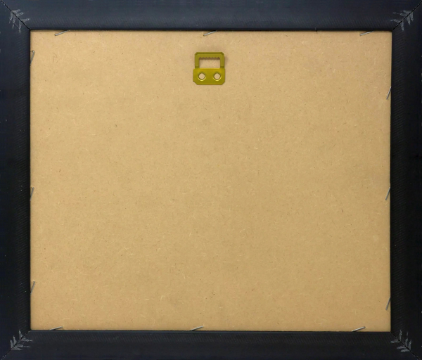
Wood effect frame, card mounted, 16x12 archival quality photo print. Overall outside dimensions 22x18 inches (58x48cm). Environmentally and ozone friendly, 40mm wide x 15mm Polycore® moulding has the look of real wood, is durable and light and easy to hang. Biodegradable and made with non-chlorinated gases (no toxic fumes) it is efficient; producing 100 tons of polystyrene can save 300 tons of trees! Prints are glazed with lightweight, shatterproof, optical clarity acrylic (providing the same general protection from the environment as glass). The back is stapled hardboard with a sawtooth hanger attached. Note: To minimise original artwork cropping, for optimum layout, and to ensure print is secure, the visible print may be marginally smaller
Contemporary Framed and Mounted Prints - Professionally Made and Ready to Hang
Estimated Image Size (if not cropped) is 22.3cm x 39.6cm (8.8" x 15.6")
Estimated Product Size is 47.8cm x 57.9cm (18.8" x 22.8")
These are individually made so all sizes are approximate
Artwork printed orientated as per the preview above, with portrait (vertical) orientation to match the source image.
FEATURES IN THESE COLLECTIONS
> Arts
> Art Movements
> Related Images
> Europe
> United Kingdom
> Scotland
> Falkirk
> Bo'ness
EDITORS COMMENTS
This artwork, titled "Brain motor cortex pathways" offers a mesmerizing glimpse into the intricate neural connections within the human brain. The print showcases a sectioned human brain, brainstem, and spinal cord, revealing the complex network of red and blue neural pathways originating from the motor cortex. These extrapyramidal pathways play a crucial role in controlling involuntary movements and coordinating muscle actions. The illustration highlights two specific tracts: the rubrospinal tract and reticulospinal tract. These pathways work harmoniously to ensure smooth movements throughout our body. The image's white background accentuates the vibrant colors of these neural pathways against an anatomical context. It is fascinating to observe how different sections of the brain are represented in this artwork – from vertical (coronal) sections at various levels such as midbrain, pons, medulla oblongata, to horizontal sections at different levels including spinal cord. By exploring this visual representation of our neurological system's complexity, we gain insight into how our brains function on both physiological and anatomical levels. This print serves as a reminder of the incredible intricacies that enable us to move effortlessly through life. Created by BO VEISLAND for Science Photo Library, this artwork combines scientific accuracy with artistic flair. Its inclusion of key structures like substantia nigra, cerebral cortex, corticopontine tract further enriches its educational value while maintaining aesthetic appeal.
MADE IN THE UK
Safe Shipping with 30 Day Money Back Guarantee
FREE PERSONALISATION*
We are proud to offer a range of customisation features including Personalised Captions, Color Filters and Picture Zoom Tools
SECURE PAYMENTS
We happily accept a wide range of payment options so you can pay for the things you need in the way that is most convenient for you
* Options may vary by product and licensing agreement. Zoomed Pictures can be adjusted in the Basket.