
Axial Collection
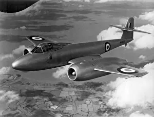
"Exploring the Axial: From Artwork to Anatomy and Aerospace" The Gloster Meteor F8 WA820 takes flight, showcasing the power propulsion


All Professionally Made to Order for Quick Shipping
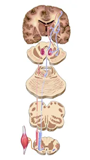
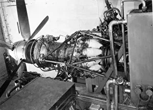
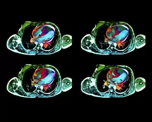
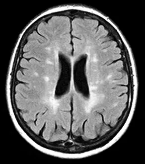
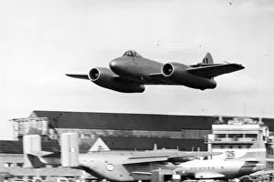
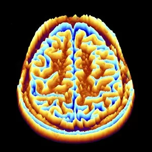
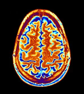
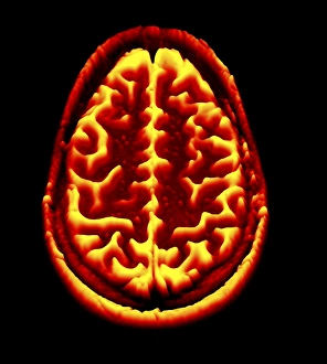
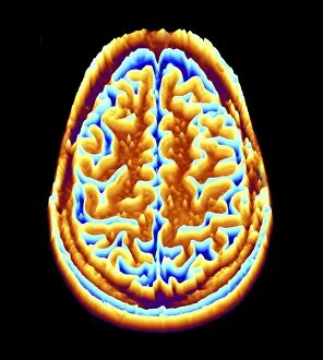
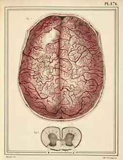
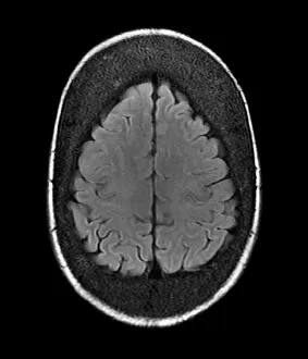
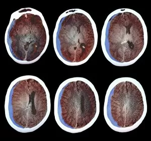
"Exploring the Axial: From Artwork to Anatomy and Aerospace" The Gloster Meteor F8 WA820 takes flight, showcasing the power propulsion. Delving into medical science, male groin arteries reveal intricate axial pathways. An 1825 artwork unveils the beauty of brain motor cortex pathways in mesmerizing detail (C016 / 6532). Frank T Verity's architectural litho captures the grandeur structures in design. Journeying back to 1878, an engraving unravels the mysteries of the axial skeleton anatomy. Witnessing innovation, Rolls Royce RB39 Clyde axial turboprop undergoes testing on a specialized testbed. Medical imaging comes alive with stroke detection through MRI and 3D CT scans (C016 / 6419). Unveiling cardiac lymphoma through detailed MRI scans that highlight its impact on axially connected organs. Exploring uterine fibroids using advanced MRI scanning techniques reveals their location within the uterus (C018 / 0466). Marvel at Gloster F9/40M DG204/G powered by two Metropolitan Vickers engines - a testament to axial engineering prowess. 11 &12: Captivating our attention once again, witness the awe-inspiring Gloster Meteor F8 WA820 as it graces us twice. In this captivating journey through various fields such as aerospace engineering, medical science, architecture, and artistry; we explore different aspects of "axial. " From aircrafts defying gravity with powerful turbines to intricate anatomical pathways connecting vital organs - these glimpses remind us of how interconnected our world truly is.