
Cerebral Cortex Collection
The cerebral cortex, also known as the outer layer of the brain, is a fascinating and intricate part of our human anatomy


All Professionally Made to Order for Quick Shipping
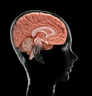
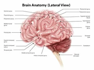
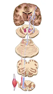
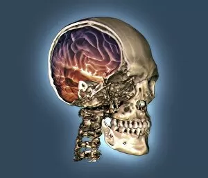
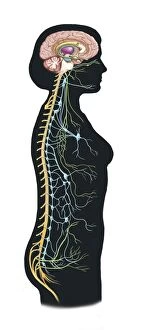
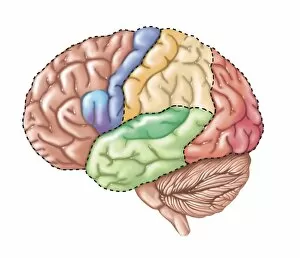
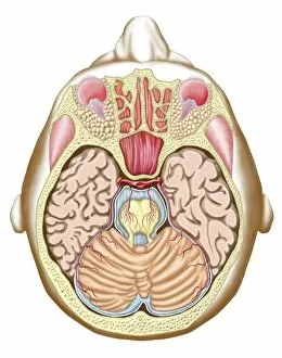
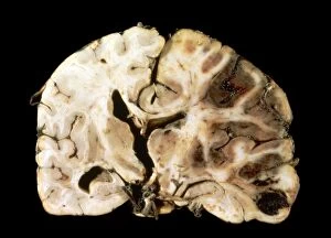
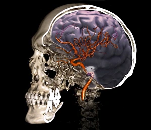
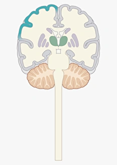
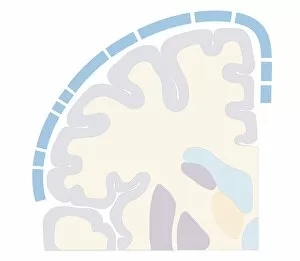
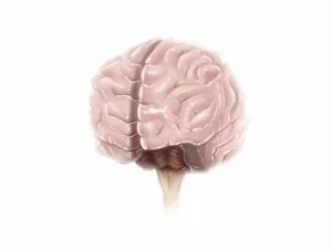
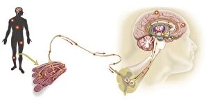
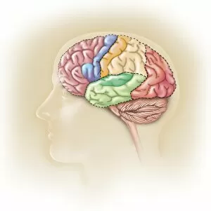
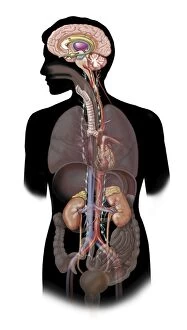
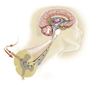
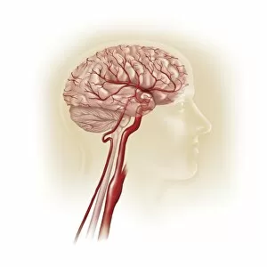
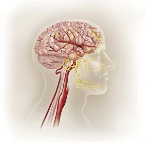
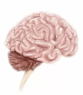
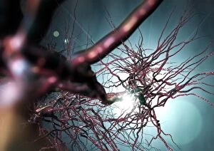
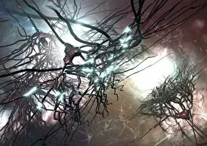
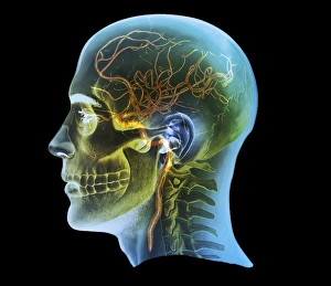
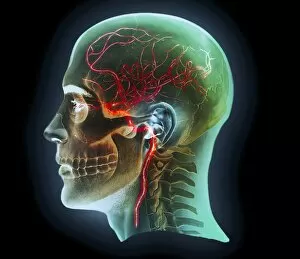
The cerebral cortex, also known as the outer layer of the brain, is a fascinating and intricate part of our human anatomy. It plays a crucial role in various functions such as perception, cognition, memory, and voluntary movement. One intriguing aspect of the cerebral cortex is its organization according to the motor homunculus model. This model depicts how different body parts are represented on specific areas of the cortex based on their importance and complexity of movements. The motor homunculus provides us with a visual representation of this intricate mapping. In addition to the motor homunculus, there is also a sensory homunculus that illustrates how different body regions are represented in terms of sensation within the cerebral cortex. Studying brain anatomy through models or MRI scans allows us to explore its complex structure further. These images provide valuable insights into understanding how nerve cells within the cerebral cortex interact and communicate with each other. Furthermore, artwork depicting brain motor cortex pathways showcases these neural connections in an artistic manner while highlighting their significance for movement control. When observing lateral views of the human brain or 3D CT scans showcasing normal skull and brain structures, we gain a deeper appreciation for our own unique neurological makeup. It's worth noting that famous psychologist Ivan Pavlov made significant contributions to our understanding of conditioning reflexes by studying animals' brains extensively. His caricature serves as a reminder of his groundbreaking research efforts. Moreover, exploring surface anatomy with labeled diagrams helps us identify key regions within the cerebral cortex responsible for various cognitive processes like language processing or decision-making. Lastly, it's essential to recognize that our autonomic nervous system and limbic system play vital roles alongside the cerebral cortex in regulating bodily functions and emotional responses throughout our entire body.