
Brain Stem Collection
The brain stem, an integral part of the anatomy of the human brain, is a complex network of pathways that plays a crucial role in our daily functioning


All Professionally Made to Order for Quick Shipping
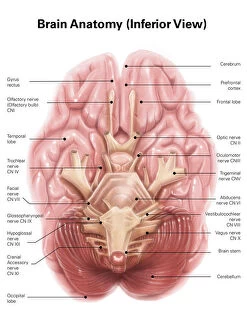
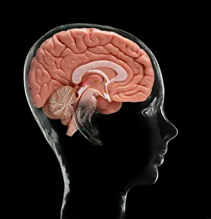
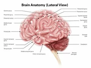
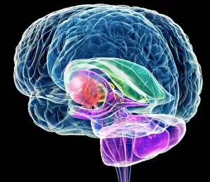
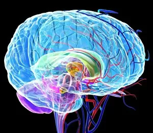
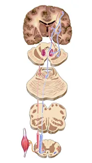
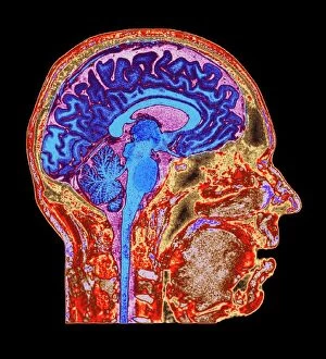
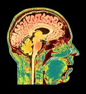
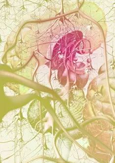
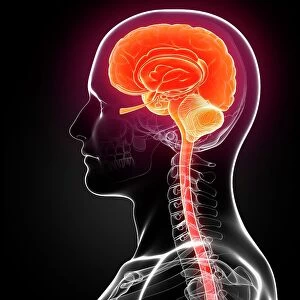
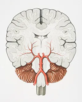
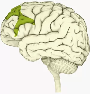
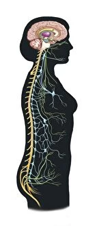
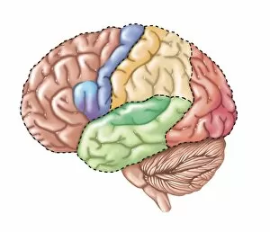
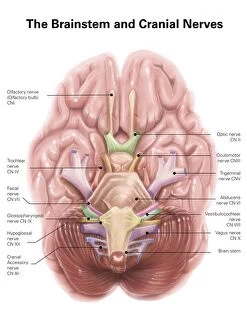
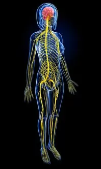
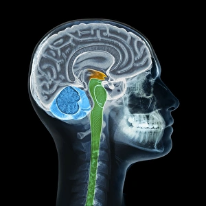
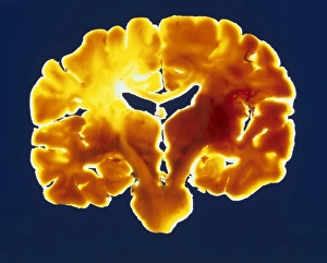
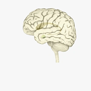
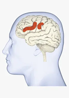
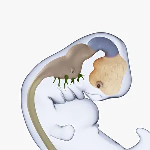
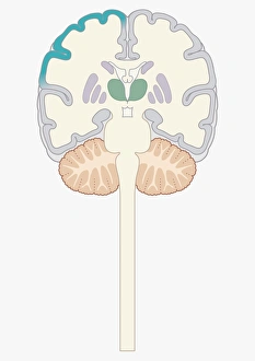
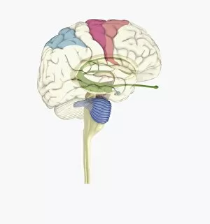
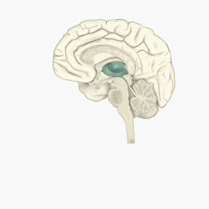
The brain stem, an integral part of the anatomy of the human brain, is a complex network of pathways that plays a crucial role in our daily functioning. When viewed from an inferior perspective, its intricate structure becomes apparent, showcasing the interconnectedness and interdependence of various regions within this vital organ. Models depicting the human brain offer a tangible representation of its complexity. From lateral views to MRI scans, these visual aids provide us with invaluable insights into the inner workings of our brains. Artwork further enhances our understanding by illustrating different aspects of brain anatomy through captivating imagery. One such artwork showcases the detailed lithograph published in 1876, which beautifully captures the intricacies and nuances of the human brain's composition. Another artwork focuses on highlighting specific pathways within the motor cortex, shedding light on how signals are transmitted throughout this essential region. MRI scans continue to revolutionize our understanding of brain health. A normal human brain scan reveals its optimal state while emphasizing areas that require attention for maintaining overall well-being. These scans serve as valuable tools in diagnosing and treating various neurological conditions. Understanding and appreciating healthy brains is crucial for advancing medical knowledge. Multiple MRI scans showcase healthy brains at different angles, providing comprehensive insights into their structures and functions. Exploring diverse perspectives such as anatomical models, artistic renderings, lithographs from centuries ago or modern-day MRI technology allows us to delve deeper into unraveling the mysteries surrounding one of nature's most remarkable creations -the intricate and awe-inspiring human brain stem