
Spinal Cord Collection
"The Spinal Cord: Unveiling the Gateway to Our Nervous System" Embarking on a journey of discovery, a full body scan reveals the intricate wonders hidden within us


All Professionally Made to Order for Quick Shipping
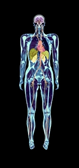
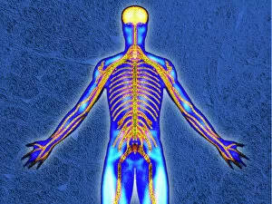
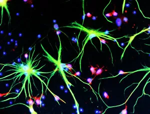
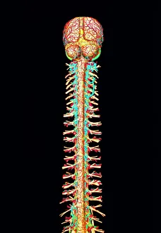
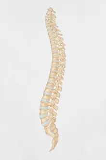
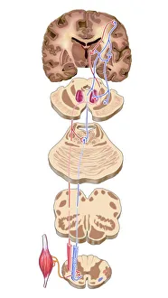
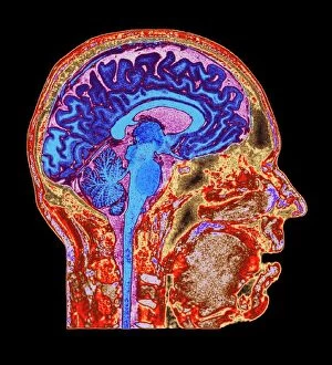
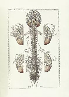
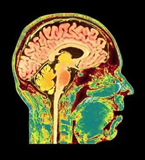
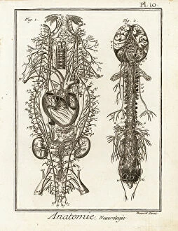
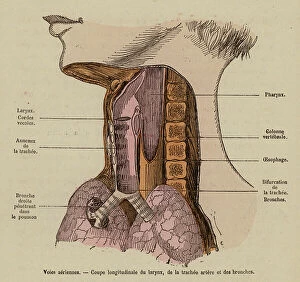
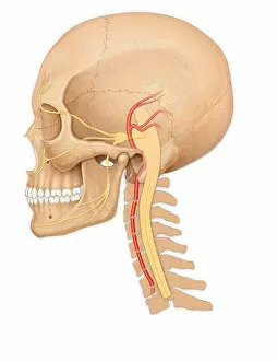
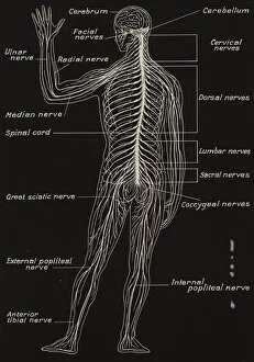
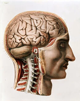
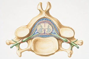
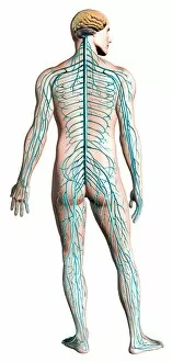
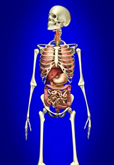
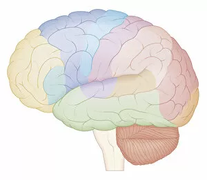
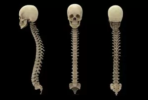
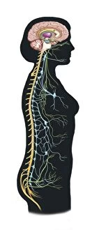
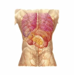
"The Spinal Cord: Unveiling the Gateway to Our Nervous System" Embarking on a journey of discovery, a full body scan reveals the intricate wonders hidden within us. An MRI scan unveils the brain's anatomy, showcasing its complexity and beauty. As our eyes wander further down, an illustration captures the human spinal cord in all its glory, intricately connected to the brain. The spinal cord serves as the central highway of our nervous system, transmitting messages through nerve cells that branch out like delicate tendrils. A diagram offers a side view of this remarkable structure - a pillar of support and communication for our entire body. Artwork by Leonardo da Vinci takes us back centuries ago when he meticulously studied and sketched pen and ink studies of the human spinal column. His dedication to understanding this vital part of our being is awe-inspiring. Moving forward in time, modern technology allows us to witness brain motor cortex pathways through mesmerizing artwork. These pathways hold secrets to how we move and interact with the world around us. A normal human brain captured by another MRI scan reminds us that every individual possesses unique neural patterns that shape their thoughts and actions. The conceptual image of a skull intertwined with the spinal cord symbolizes their inseparable connection - one cannot exist without the other. Delving deeper into history, Bartholomeo Eustachi's "The Science of Human Anatomy" showcases humanity's relentless pursuit to unravel nature's mysteries. It stands as a testament to our ceaseless curiosity about ourselves. These captivating glimpses into various aspects surrounding the spinal cord remind us just how extraordinary it truly is. From ancient sketches by da Vinci to cutting-edge imaging techniques today, we continue exploring this gateway between mind and body – forever fascinated by its profound significance in shaping who we are as humans.