
Glass Frame : Spine anatomy


![]()
Mounted Prints from Science Photo Library
Spine anatomy
Spine anatomy. Coloured light micrograph of a section through a spine, showing vertebra bone (purple), the spinal cord (yellow, upper centre), the spinal canal (white) and surrounding tissue (brown). A vertebra is a bone segment that links with other vertebra to make up the spine (backbone). The spinal canal is a hollow through which the spinal cord and spinal nerve (thick pink ring around the cord) runs. Magnification: x10 when printed 10cm wide
Science Photo Library features Science and Medical images including photos and illustrations
Media ID 6420192
© STEVE GSCHMEISSNER/SCIENCE PHOTO LIBRARY
Bones Cavity Cross Section False Colour Histological Histology Hollow Joint Slice Spinal Cord Tissue Transverse Section Tube Vertebra False Coloured Light Micrograph Light Microscope Section Sectioned Vertebrae
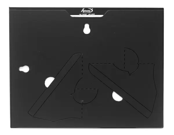
8"x6" Glass Mount
Wall mounted or free-standing, these black edged glass frames feature a smooth chamfered edge and a stylish black border (on back face of the glass). Manufactured from 4mm thick glass, Glass Mounts are a durable, professional way of displaying and protecting your prints. Your 8x6 print is slotted into the back of the frame so can easily be changed if needed.
Tempered Glass Mounts are ideal for wall display, plus the smaller sizes can also be used free-standing via an integral stand
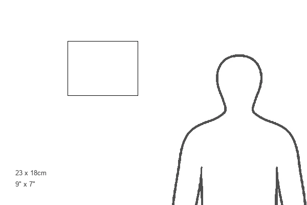
Estimated Image Size (if not cropped) is 20.3cm x 15.2cm (8" x 6")
Estimated Product Size is 22.8cm x 17.7cm (9" x 7")
These are individually made so all sizes are approximate
Artwork printed orientated as per the preview above, with landscape (horizontal) orientation to match the source image.
EDITORS COMMENTS
This print showcases the intricate anatomy of the spine in vibrant colors. The image reveals a cross-section of the spine, highlighting various essential components. The vertebrae, represented by a striking purple hue, are bone segments that form the backbone and provide structural support to our bodies. At the center of attention is the spinal cord, depicted in a radiant yellow shade. This vital part of our nervous system runs through the hollow spinal canal, which appears as an ethereal white tube. Encircling the spinal cord is a thick pink ring representing the spinal nerve. The surrounding tissue is beautifully captured in shades of brown, emphasizing its connection to this complex network within our bodies. This micrograph provides us with an up-close view of normal anatomical features and offers valuable insights into human biology. With a magnification level of x10 when printed at 10cm wide, every detail becomes apparent and mesmerizing. Whether you're studying medicine or simply fascinated by science, this image serves as a remarkable visual aid for understanding spine anatomy. Courtesy of Science Photo Library's extensive collection, this print exemplifies their commitment to providing high-quality scientific imagery for educational purposes.
MADE IN THE UK
Safe Shipping with 30 Day Money Back Guarantee
FREE PERSONALISATION*
We are proud to offer a range of customisation features including Personalised Captions, Color Filters and Picture Zoom Tools
SECURE PAYMENTS
We happily accept a wide range of payment options so you can pay for the things you need in the way that is most convenient for you
* Options may vary by product and licensing agreement. Zoomed Pictures can be adjusted in the Basket.