Fraud Blocker Skip to main content

Vertebrae Collection

"Unlocking the Secrets of Vertebrae: Exploring Chakras and the Nervous System" Delve into the intricate world of vertebrae

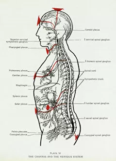
Background imageVertebrae Collection: Chakras and Nervous System

Chakras and Nervous System
How the chakras interact with the nervous system. Date: 1927

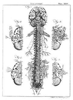
Background imageVertebrae Collection: Diagram of the human brain and spinal column

Diagram of the human brain and spinal column
A diagram of the brain and spinal column, including the vertebrae

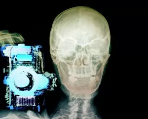
Background imageVertebrae Collection: Person with a camera, X-ray

Person with a camera, X-ray
Person with a camera. Coloured, frontal X-ray of a person holding a camera (blue, left). The persons skull (centre) and neck bones (vertebrae, lower centre) can be seen

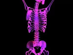
Background imageVertebrae Collection: Upper body skeleton, computer artwork

Upper body skeleton, computer artwork
Upper body skeleton. Computer artwork of a human skeleton showing the bones of the upper body. At the top is the base of the skull and the jaw bone

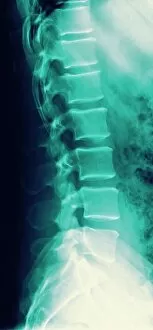
Background imageVertebrae Collection: Normal spine, X-ray

Normal spine, X-ray
Normal spine. Coloured profile X-ray showing bones of the spine (vertebrae, centre). The front of the body is at right. The ribs (upper right) and the pelvis (bottom) are also seen

Background imageVertebrae Collection: Normal neck, X-ray

Normal neck, X-ray
Noraml neck. X-ray of the healthy cervical (neck) spine of a 29 year old male patient

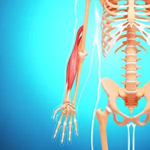
Background imageVertebrae Collection: Human arm musculature, artwork F007 / 1810

Human arm musculature, artwork F007 / 1810
Human arm musculature, computer artwork

Background imageVertebrae Collection: Dog skeleton, artwork F006 / 2267

Dog skeleton, artwork F006 / 2267
Dog skeleton, computer artwork

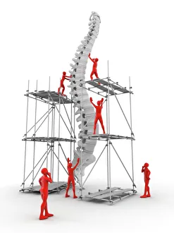
Background imageVertebrae Collection: Spine with workers, spine repair F007 / 9907

Spine with workers, spine repair F007 / 9907
Conceptual computer artwork depicting a spine being repaired

Background imageVertebrae Collection: Plate 19. Mantells Geology of Sussex

Plate 19. Mantells Geology of Sussex
Plate 19 from Illustrations of the Geology of Sussex, 1827 by G. A. Mantell

Background imageVertebrae Collection: Neck, side view, coloured X-ray

Neck, side view, coloured X-ray
Neck from the side, coloured X-ray. The front of the body is at right. The spine runs down centre. It is made up of block-like bones called vertebrae

Background imageVertebrae Collection: Slipped disc

Slipped disc. Spinal model demonstrating a slipped intervertebral disc. The slipped disc is the small bulge (red, centre), near the base of the spine

Background imageVertebrae Collection: Neck pain, conceptual artwork

Neck pain, conceptual artwork

Background imageVertebrae Collection: Normal torso, MRI scan

Normal torso, MRI scan
Normal torso, coloured profile (sagittal) magnetic resonance imaging (MRI) scan. The front of the body is at left. The spinal bones (vertebrae)

Background imageVertebrae Collection: Slipped intervertebral disc, artwork

Slipped intervertebral disc, artwork
Slipped intervertebral disc, computer artwork. Intervertebral discs (red) are pads of cartilage between the bones (vertebrae) of the spine

Background imageVertebrae Collection: Inflamed spinal discs, MRI scan

Inflamed spinal discs, MRI scan
Inflamed spinal discs. Coloured magnetic resonance imaging (MRI) scan of a side view of the spine of a patient with discitis. The front of the body is at left, the skull is at top

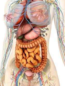
Background imageVertebrae Collection: Female body showing digestive and circulatory system

Female body showing digestive and circulatory system
Medical illustration of female body showing digestive and circulatory system, perspective view

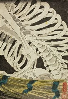
Background imageVertebrae Collection: Mitsukuni and the Skeleton Specter (image 3 of 3), mid 1840s. Creator: Utagawa Kuniyoshi

Mitsukuni and the Skeleton Specter (image 3 of 3), mid 1840s. Creator: Utagawa Kuniyoshi
Mitsukuni and the Skeleton Specter (image 3 of 3), mid 1840s

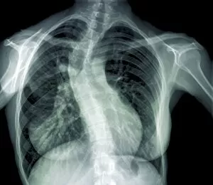
Background imageVertebrae Collection: Scoliosis of the spine, X-ray

Scoliosis of the spine, X-ray
Scoliosis of the spine. Coloured X-ray of the chest of a 31 year old patient with scoliosis (sideways curvature) of the spine

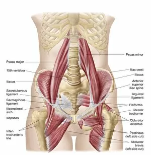
Background imageVertebrae Collection: Anatomy of iliopsoa, also known as the dorsal hip muscles

Anatomy of iliopsoa, also known as the dorsal hip muscles
Anatomy of iliopsoa, often referred to as the dorsal hip muscles. These muscles are distinct in the abdomen

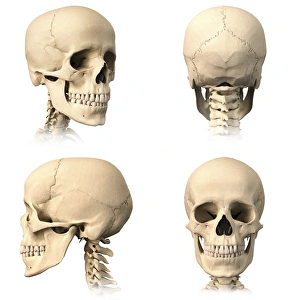
Background imageVertebrae Collection: Anatomy of human skull from different angles

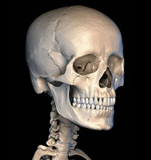
Anatomy of human skull from different angles

Background imageVertebrae Collection: Study of a Fowl Head and Neck, Lateral View, from A Comparative Anatomical Exposition of

Study of a Fowl Head and Neck, Lateral View, from A Comparative Anatomical Exposition of the Structure of the Human
XYC262893 Study of a Fowl Head and Neck, Lateral View, from A Comparative Anatomical Exposition of the Structure of the Human Body with that of a Tiger and a Common Fowl

Background imageVertebrae Collection: Human skull, illustration

Human skull, illustration
Human skull, computer illustration

Background imageVertebrae Collection: California Usa America American Death Valley

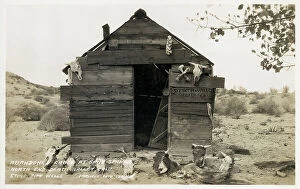
California Usa America American Death Valley
california, usa, america, american, death, valley, abandoned, cabin, shed, wood, wooden, makeshift, building, buildings, ramshackle, animal, skulls, skull, bone, bones, sand, springs, north, end

Background imageVertebrae Collection: DDE-90034363

DDE-90034363
Norway, Spitsbergen, Sallyhammna. Vertebrae of a dead fin whale (Balaenoptera physalus) lies on a snow-covered beach one year after its death, glacier in distance. Date: 14/04/2010

Background imageVertebrae Collection: DDE-90034362

DDE-90034362
Norway, Spitsbergen, Sallyhammna. Vertebrae of a dead fin whale (Balaenoptera physalus) lies on a beach one year after its death, glacier in distance. Date: 14/04/2010

Background imageVertebrae Collection: DDE-90034318

DDE-90034318
Norway, Spitsbergen, Fuglefjorden. Polar bear (Ursus maritimus) adult feeds on the carcass of a bearded seal (Erignathus barbatus) pup catch. Date: 14/04/2010

Background imageVertebrae Collection: DDE-90034319

DDE-90034319
Norway, Spitsbergen, Fuglefjorden. Polar bear (Ursus maritimus) adult feeds on the carcass of a bearded seal (Erignathus barbatus) pup catch. Date: 14/04/2010

Background imageVertebrae Collection: DDE-90034317

DDE-90034317
Norway, Spitsbergen, Fuglefjorden. Polar bear (Ursus maritimus) adult feeds on the carcass of a bearded seal (Erignathus barbatus) pup catch. Date: 14/04/2010

Background imageVertebrae Collection: DDE-90034316

DDE-90034316
Norway, Spitsbergen, Fuglefjorden. Polar bear (Ursus maritimus) adult feeds on the carcass of a bearded seal (Erignathus barbatus) pup catch. Date: 14/04/2010

Background imageVertebrae Collection: DDE-90033904

DDE-90033904
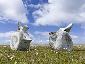
Norway, Svalbard, Storfjorden Fjord, Whale vertebrae standing on tundra on summer afternoon in Tusenoyane Archipelago Date: 17/07/2007

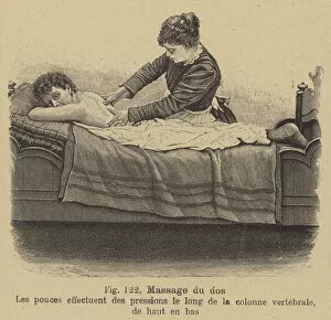
Background imageVertebrae Collection: Back massage (engraving)

Back massage (engraving)
3097159 Back massage (engraving) by German School, (19th century); Private Collection; (add.info.: Back massage. Using the thumbs to massage the spinal column from top to bottom)

Background imageVertebrae Collection: Sceleta Strenis Lacertinae et Protei Mexicani, plate XIV

Sceleta Strenis Lacertinae et Protei Mexicani, plate XIV
HMB321596 Sceleta Strenis Lacertinae et Protei Mexicani, plate XIV, Osteology of the sirenomelia and axolotl (litho) by Humboldt, Friedrich Alexander

Background imageVertebrae Collection: Remains of an Extinct Animal found in the Isle of Wight (engraving)

Remains of an Extinct Animal found in the Isle of Wight (engraving)
1610597 Remains of an Extinct Animal found in the Isle of Wight (engraving) by English School, (19th century); Private Collection; (add.info.: Remains of an Extinct Animal found in the Isle of Wight)

Background imageVertebrae Collection: The Sceleton of the Backe, plate from The Historie of Man

The Sceleton of the Backe, plate from The Historie of Man
AFC116188 The Sceleton of the Backe, plate from The Historie of Man published by John Banister (1540-1610), 1578 (engraving) by English School

Background imageVertebrae Collection: Whale vertebrae on Bleaker Island. South America, FAlkland Islands, January

Whale vertebrae on Bleaker Island. South America, FAlkland Islands, January

Background imageVertebrae Collection: Human skeleton, rib cage and shoulder blades, high angle view

Human skeleton, rib cage and shoulder blades, high angle view

Background imageVertebrae Collection: Anatomy of human skull, rear view

Anatomy of human skull, rear view

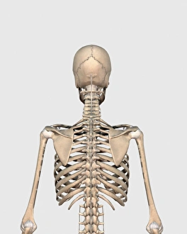
Background imageVertebrae Collection: Rear view of human skeletal system showing upper back

Rear view of human skeletal system showing upper back

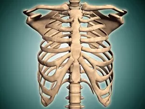
Background imageVertebrae Collection: Close-up view of human rib cage

Close-up view of human rib cage

Background imageVertebrae Collection: Anterior view of human pelvis, with labels

Anterior view of human pelvis, with labels

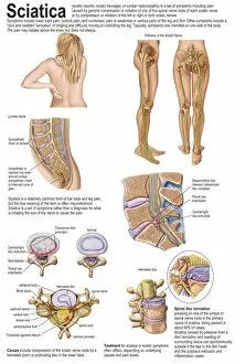
Background imageVertebrae Collection: Medical chart showing the signs and symptoms of sciatica

Medical chart showing the signs and symptoms of sciatica

Background imageVertebrae Collection: Picture No. 10875835

Picture No. 10875835
Human skeleton - body structure - skull and Vertebrae side view Date:

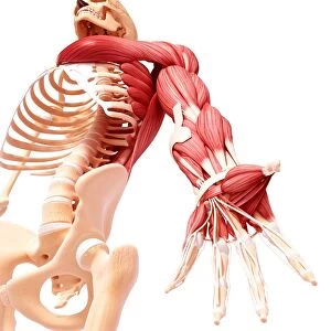
Background imageVertebrae Collection: Human arm musculature, artwork F007 / 1379

Human arm musculature, artwork F007 / 1379
Human arm musculature, computer artwork

Background imageVertebrae Collection: Normal head and neck, MRI and 3D CT scans C016 / 6337

Normal head and neck, MRI and 3D CT scans C016 / 6337
Normal head. Coloured composite image of a magnetic resonance imaging (MRI) scan of the brain and a 3D computed tomography (CT) scan of the head and neck of a 35 year old patient

Background imageVertebrae Collection: Chest bones

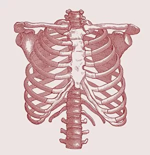
Chest bones. Historical artwork of the bones of the human chest. The breastbone (upper centre) at the front of the chest supports 12 pairs of ribs that circle the chest and connect to the backbone

Background imageVertebrae Collection: Red deer skeleton

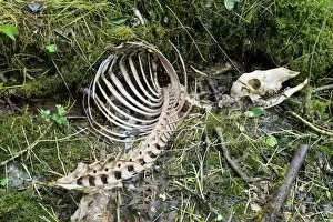
Red deer skeleton. Part of the skeleton of a red deer (Cervus elaphus), found in a wood near a ditch

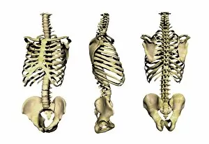
Background imageVertebrae Collection: Human skeleton anatomy, artwork

Human skeleton anatomy, artwork
Human skeleton anatomy, computer artwork. Three views of the bones of the human torso, seen from an oblique frontal view (left), from the side (centre) and from the front (right)


All Professionally Made to Order for Quick Shipping

"Unlocking the Secrets of Vertebrae: Exploring Chakras and the Nervous System" Delve into the intricate world of vertebrae, where chakras and the nervous system intertwine like a beautifully orchestrated symphony. A diagram showcasing the human brain and spinal column reveals the remarkable connection between our thoughts and physical well-being. With a camera in hand, capture an X-ray image that unveils both the fragility and strength of our vertebral structure. The upper body skeleton depicted in computer artwork serves as a reminder of how these bones provide support for every movement we make. Observe a normal neck through an X-ray lens, appreciating its graceful alignment that allows us to turn our heads with ease. An X-ray image displaying a normal spine showcases its incredible flexibility while maintaining stability throughout our lives. Through captivating artwork, explore the complexity of human arm musculature intertwined with vertebrae – truly a marvel of nature's design (F007 / 1810). Even animals possess this fascinating skeletal structure; admire a dog skeleton captured in detailed artwork (F006 / 2267). Witness spine repair in action as workers diligently restore damaged vertebrae – highlighting humanity's dedication to healing (F007 / 9907). Plate 19 from Mantell's Geology of Sussex offers insight into how geological forces have shaped even our own vertebral history. A colored X-ray provides a side view glimpse into the delicate intricacies within one's neck – reminding us to cherish this vital part of ourselves. Sometimes, challenges arise such as slipped discs - but modern medicine continues to innovate ways to heal and alleviate discomfort. Intriguingly complex yet undeniably essential, vertebrae serve as both guardians protecting our nervous system and conduits connecting mind and body.