
Vertebra Collection
"Unveiling the Backbone of Life: Exploring the Fascinating World of Vertebrae" A glimpse into history reveals a horse's skull


All Professionally Made to Order for Quick Shipping
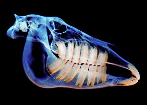
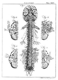
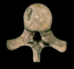
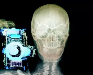
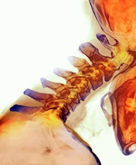
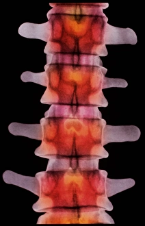
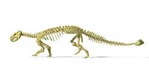
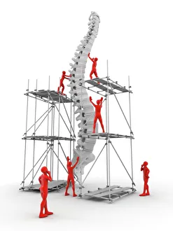
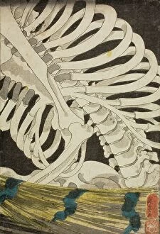
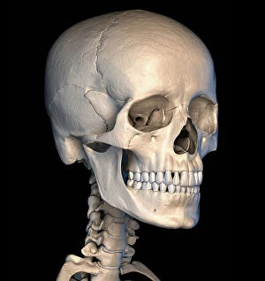
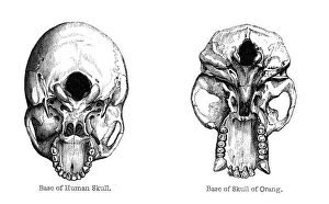
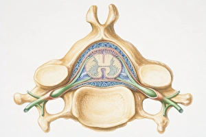
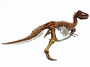
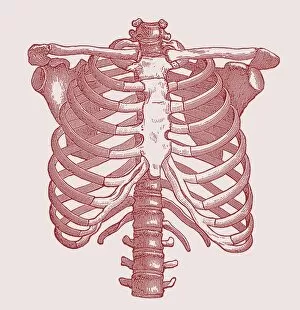
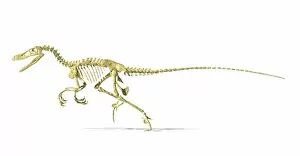
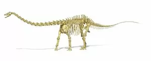
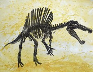
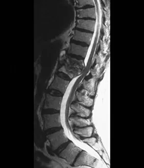
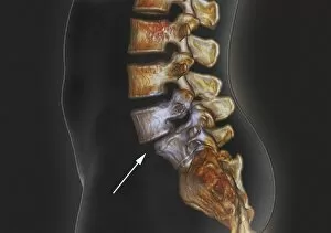
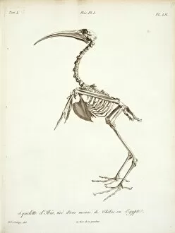
"Unveiling the Backbone of Life: Exploring the Fascinating World of Vertebrae" A glimpse into history reveals a horse's skull, showcasing the intricate vertebrae that form its backbone. Delving deeper, a diagram illustrates the human brain and spinal column, highlighting how vital vertebrae are to our nervous system. With a camera in hand, an individual captures an X-ray image, unveiling the hidden secrets within vertebral structures. Journeying back in time, we encounter a Liopleurodon vertebra—a relic from prehistoric oceans—reminding us of ancient creatures' remarkable skeletal systems. Witnessing neck vertebrae extended through an X-ray image showcases their flexibility and adaptability for various movements. An artistically colored X-ray depicts lumbar vertebrae—the foundation of our spine—revealing their strength and interconnectedness. Examining a normal neck through an X-ray unveils its structural integrity—an essential component for supporting our head and facilitating movement. Immerse yourself in paleontological wonders with a 3D rendering of an Ankylosaurus dinosaur skeleton, emphasizing its robust vertebral framework. Peering inside another 3D rendering—this time featuring a Tyrannosaurus Rex dinosaur skeleton—we marvel at how these extinct giants relied on their powerful spines for survival. Unearthed from centuries past is an illustration depicting the nervous system as understood during the 18th century—a testament to humanity's evolving knowledge about vertebral significance over time. In modern times, workers collaborate to repair damaged spines—an inspiring reminder of medical advancements aimed at restoring mobility and quality of life for individuals facing spinal issues (Spine with workers; spine repair F007 / 9907).