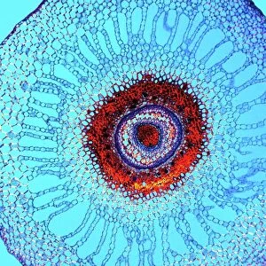
Fern rhizome, light micrograph

![]()
Wall Art and Photo Gifts from Science Photo Library
Fern rhizome, light micrograph
Fern rhizome, light micrograph. Transverse section through the center of a rhizome from the Killarney fern (Trichomanes speciosum). Thick-walled sclerenchyma cells (black-red) surround the central vascular cylinder (stele). From the outside of the stele inwards are four thin layers: the endodermis (red), the pericycle, and two layers of phloem. In the centre are the large thick-walled cells of the xylem. Magnification: x100 when printed at 10 centimetres wide
Science Photo Library features Science and Medical images including photos and illustrations
Media ID 6307831
© DR KEITH WHEELER/SCIENCE PHOTO LIBRARY
Cellular Circle Circular Cross Section Endodermis Fern Ferns Internal Structure Part Parts Pericycle Phloem Plant Anatomy Pteridophyte Pteridophytes Rhizome Round Sclerenchyma Stele Structural Tissue Transverse Vascular Cylinder Xylem Cells Light Micrograph Light Microscope Section Sectioned
EDITORS COMMENTS
This print showcases the intricate beauty of a fern rhizome, as seen through a light micrograph. The image captures a transverse section through the center of a rhizome from the Killarney fern, scientifically known as Trichomanes speciosum. The visual is composed of various elements that highlight the internal structure of this botanical wonder. Surrounding the central vascular cylinder, we can observe thick-walled sclerenchyma cells in striking black-red hues. Moving inward from the stele are four distinct layers: starting with the vibrant red endodermis, followed by the pericycle and two layers of phloem. At its core lies an awe-inspiring arrangement of large thick-walled xylem cells that form a circular pattern within this circular cross-section. When printed at 10 centimeters wide, this magnified view offers an astonishing level of detail at 100x magnification. This remarkable photograph not only celebrates nature's intricate designs but also serves as a valuable resource for botany enthusiasts and researchers alike. It provides insight into plant anatomy, specifically highlighting cellular structures and their roles within ferns' internal systems. With its scientific significance and aesthetic appeal, this print is truly an exquisite representation of how artistry and biology intertwine to create breathtaking visuals in our natural world.
MADE IN THE UK
Safe Shipping with 30 Day Money Back Guarantee
FREE PERSONALISATION*
We are proud to offer a range of customisation features including Personalised Captions, Color Filters and Picture Zoom Tools
SECURE PAYMENTS
We happily accept a wide range of payment options so you can pay for the things you need in the way that is most convenient for you
* Options may vary by product and licensing agreement. Zoomed Pictures can be adjusted in the Basket.