
Endodermis Collection
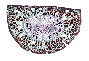
The endodermis, a vital layer within plant roots and stems, plays a crucial role in regulating the movement of water and nutrients


All Professionally Made to Order for Quick Shipping
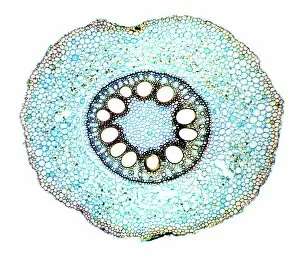
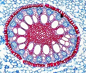
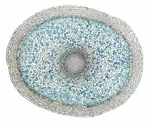
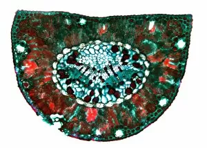
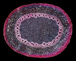
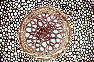
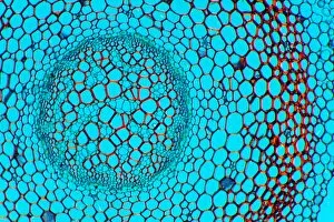
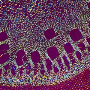
The endodermis, a vital layer within plant roots and stems, plays a crucial role in regulating the movement of water and nutrients. Underneath the epidermis lies this specialized tissue that acts as a barrier for selective nutrient absorption. Examining the endodermis under scanning electron microscopy (SEM) reveals its intricate structure. Vascular bundles can be observed running through this layer, transporting water and minerals throughout the plant. In maize roots, light micrographs showcase the distinct arrangement of cells within the endodermis, highlighting its importance in maintaining proper nutrient balance. Further exploration into different plant species uncovers fascinating variations in endodermal structures. Iris root exhibits an organized pattern of cells while Acorus calamus rhizome displays a more irregular arrangement. Light micrographs of Dendrobium orchid roots reveal unique cell shapes and sizes within their endodermis layers. Notably, Dendrobium orchids exhibit multiple light micrographs showcasing their complex root systems. These images demonstrate how the endodermis adapts to support these beautiful plants' growth and survival. Additionally, Smilax root presents an intriguing view with its densely packed cells forming a protective shield around vascular tissues. Another glimpse at Acorus calamus rhizome highlights how even closely related plants can have distinct characteristics within their endodermal layers. Lastly, exploring Clubmoss stem provides insight into how this ancient plant group's endodermis has evolved over time to suit its specific needs. In summary, studying various specimens through SEM or light micrography allows us to appreciate the diverse structures present in different types of endoderms across plant species. Understanding these intricacies helps scientists unravel nature's secrets behind efficient nutrient transport mechanisms essential for healthy growth and development.