
Pericycle Collection
The pericycle: a hidden world beneath the surface. 🌱✨ Delve into the intricate beauty of plant life with these stunning light micrographs


All Professionally Made to Order for Quick Shipping
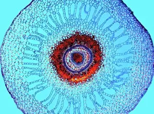
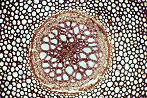
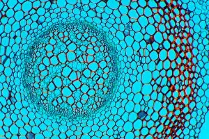
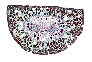
The pericycle: a hidden world beneath the surface. 🌱✨ Delve into the intricate beauty of plant life with these stunning light micrographs. From water fern rhizomes to wheat leaves, each image reveals the fascinating structures within. In the depths of a water fern rhizome, we witness an intricate network of cells that support growth and nutrient transport. The Acorus calamus rhizome showcases its unique arrangement, providing stability and nourishment for this remarkable plant. Dendrobium orchid roots captivate us with their delicate yet resilient nature. These light micrographs unveil a complex system designed for absorption and anchorage. It's truly astonishing how such elegance can be found in every root strand. As we explore further, we encounter the Smilax root, revealing its strength and adaptability through its cellular composition. This tenacious root demonstrates nature's ability to thrive even in challenging environments. Moving above ground, we observe wheat leaves in all their glory – a symphony of chlorophyll-filled cells working tirelessly to convert sunlight into energy. These light micrographs showcase the incredible efficiency of photosynthesis at work. Returning underground once more, Dendrobium orchid roots reappear on our journey – reminding us of their importance as they provide sustenance for these magnificent flowers above ground. Clubmoss stems take center stage next with their intricate vascular systems that enable them to grow tall while maintaining structural integrity. Nature's engineering marvels never cease to amaze. Lastly, sage stems reveal themselves through these captivating light micrographs – showcasing both strength and flexibility as they support foliage above while absorbing nutrients below. The pericycle invites us into a realm where beauty meets functionality; where microscopic wonders shape our natural world. Let these mesmerizing images remind us to appreciate not only what lies before our eyes but also what lies beneath - unseen but essential for life itself.