
Transverse Collection
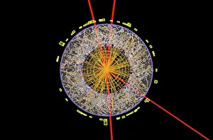
"Exploring the Transverse World: From Subatomic Particles to Botanical Wonders" Unveiling the Secrets: The Higgs Boson Event captured by the ATLAS detector C013/6892


All Professionally Made to Order for Quick Shipping
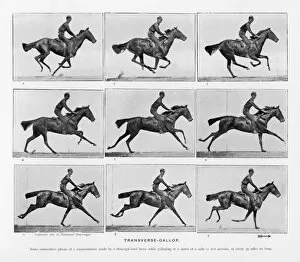
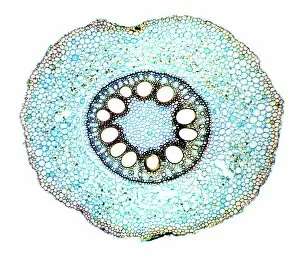
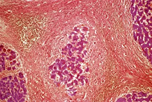
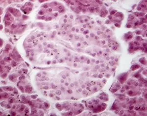
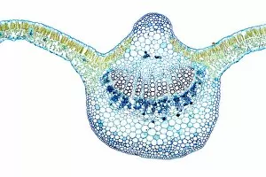
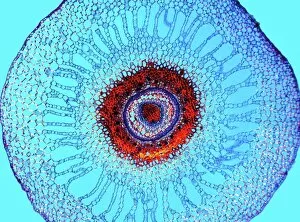
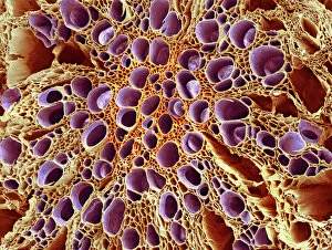
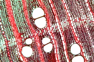
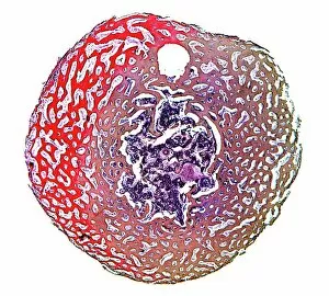
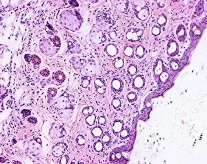
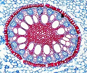
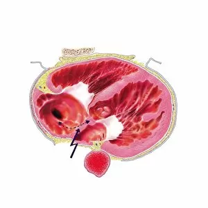
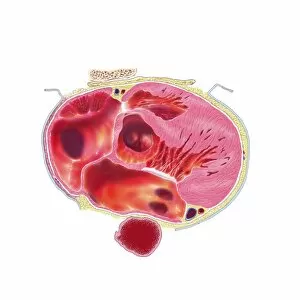
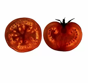
"Exploring the Transverse World: From Subatomic Particles to Botanical Wonders" Unveiling the Secrets: The Higgs Boson Event captured by the ATLAS detector C013/6892. Frozen in Time: Muybridge's iconic study of Horses Galloping reveals their transverse motion. Nature's Engineering Marvels: Witnessing the intricate Vascular Bundle through a SEM image. Beneath the Surface: A closer look at Compact Bone structure under a light micrograph. Revving Up History: Mechanics examine Bugatti Type 251 engine during the 1956 French Grand Prix. Rooted in Life: Maize Root depicted in stunning detail under a light micrograph. Battling Disease: Light micrograph showcases Liver Tissue Cirrhosis, revealing its transverse impact on health. Pancreatic Powerhouse: Islet of Langerhans illuminated through a captivating light micrograph. Hidden Depths Below Ground: Exploring Water Fern Rhizome's intricate network using a light micrograph technique. Tall and Strong Guardians of Nature - Pine Tree Stem revealed through an enchanting light micrograph image Xylem Tissue Exposed. Delicate structures captured with precision using SEM technology Wood Underneath Our Feet.