Greetings Card : Pancreas cells, SEM


![]()
Cards from Science Photo Library
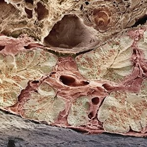
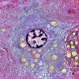
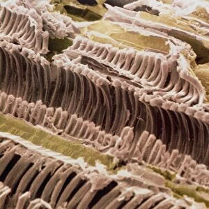
Pancreas cells, SEM
Pancreas cells. Coloured scanning electron micrograph (SEM) of acinar (exocrine) pancreatic cells. Acinar cells produce and excrete digestive enzymes to the small intestine, via the pancreatic ducts. The enzymes are excreted in vesicles known as zymogen granules (red). The enzymes are activated in the small intestine and aid the breakdown of carbohydrates, fats and proteins. Magnification: x330 when printed 10 centimetres wide
Science Photo Library features Science and Medical images including photos and illustrations
Media ID 6450003
© STEVE GSCHMEISSNER/SCIENCE PHOTO LIBRARY
Digestive Enzymatic Enzyme Enzymes Excretory Exocrine False Colour Fracture Fractured Granules Histological Histology Pancreas Pancreatic Physiological Physiology Storage Transport Vesicle Vesicles Zymogen Granule False Coloured Section Sectioned
Greetings Card (A5)
Brighten someone's day with a unique and educational greeting card from Media Storehouse. This captivating design features a colorful Scanning Electron Micrograph (SEM) image of pancreas cells by Science Photo Library. Delve into the wonders of biology as you explore the intricate structure of acinar pancreatic cells, which produce and secrete digestive enzymes to the small intestine. A perfect card for science enthusiasts, students, or anyone who appreciates the beauty of the natural world. Add a thoughtful message and send a dose of knowledge and inspiration with every greeting.
Create your own greetings card. Size when folded is A5 (14.5x21cm or 5.7x8.3 inches)
Greetings Cards suitable for Birthdays, Weddings, Anniversaries, Graduations, Thank You and much more
Estimated Image Size (if not cropped) is 21cm x 14.5cm (8.3" x 5.7")
Estimated Product Size is 21cm x 29cm (8.3" x 11.4")
These are individually made so all sizes are approximate
Artwork printed orientated as per the preview above, with landscape (horizontal) orientation to match the source image.
EDITORS COMMENTS
This print showcases the intricate beauty of pancreas cells, captured through a scanning electron microscope (SEM). The image is colored to highlight the various components and structures within these exocrine pancreatic cells. Acinar cells, depicted here in stunning detail, play a crucial role in our digestive system by producing and releasing digestive enzymes into the small intestine via specialized ducts. The zymogen granules, seen as vibrant red vesicles within the image, contain these powerful enzymes that aid in breaking down carbohydrates, fats, and proteins during digestion. Once activated in the small intestine, they contribute to our body's physiological processes. With a magnification of x330 when printed at 10 centimeters wide, this photograph allows us to appreciate the complexity and organization of these vital cellular structures. It offers an intriguing glimpse into the microscopic world that underlies our biological functions. Whether you are studying biology or simply fascinated by human anatomy and physiology, this visually striking SEM image provides both scientific insight and aesthetic appeal. Its false-colored presentation adds an artistic touch while maintaining its scientific integrity. This print from Science Photo Library serves as a reminder of how even at such minuscule scales, nature's wonders never cease to amaze us.
MADE IN THE UK
Safe Shipping with 30 Day Money Back Guarantee
FREE PERSONALISATION*
We are proud to offer a range of customisation features including Personalised Captions, Color Filters and Picture Zoom Tools
SECURE PAYMENTS
We happily accept a wide range of payment options so you can pay for the things you need in the way that is most convenient for you
* Options may vary by product and licensing agreement. Zoomed Pictures can be adjusted in the Basket.