Fraud Blocker Skip to main content

Pancreatic Collection

The pancreatic organ, located in the abdomen, is a fascinating and intricate part of our anatomy

Background imagePancreatic Collection: Pancreas anatomy, artwork

Pancreas anatomy, artwork
Pancreas anatomy. Cut-away computer artwork of the pancreas, which secretes digestive enzymes and the hormones insulin and glucagon, which regulate blood sugar levels

Background imagePancreatic Collection: Islet of Langerhans, light micrograph

Islet of Langerhans, light micrograph
Islet of Langerhans. Light micrograph of a section through an islet of Langerhans (centre) in the pancreas. This structure is composed of clumps of secretory cells

Background imagePancreatic Collection: Pancreatic islet of Langerhans

Pancreatic islet of Langerhans
Islet of Langerhans. Light micrograph of a section through an islet of Langerhans (pale, centre) in pancreas tissue. This clump of secretory cells forms part of the endocrine system of the body

Background imagePancreatic Collection: The Pancreas (colour litho)

The Pancreas (colour litho)
CHT235086 The Pancreas (colour litho) by Aselli, Gasparo (1581-1626); Bibliotheque de la Faculte de Medecine, Paris, France; eArchives Charmet; Italian, out of copyright

Background imagePancreatic Collection: Pancreatic acinar cell

Pancreatic acinar cell. Transmission electron micrograph (TEM) of a section through an enzyme-secreting acinar cell in the human pancreas, showing part of the nucleus (round, far left)

Background imagePancreatic Collection: Ghrelin hormone molecule

Ghrelin hormone molecule. Computer model showing the crystal structure of the human hormone ghrelin. The crystal structure consists of both the secondary structure

Background imagePancreatic Collection: Insulin molecule C014 / 2121

Insulin molecule C014 / 2121
Insulin molecule. Molecular module of insulin showing its secondary structure. Insulin is a hormone produced by the pancreas

Background imagePancreatic Collection: Insulin molecule F006 / 9761

Insulin molecule F006 / 9761
Insulin molecule. Molecular model of the hormone insulin from a pig. Insulin consists of two peptide chains, A and B, which are linked by disulphide bridges

Background imagePancreatic Collection: Trypsin molecule F006 / 9634

Trypsin molecule F006 / 9634
Trypsin molecule. Molecular model of the digestive protease enzyme trypsin. Trypsin is released by the pancreas to break down proteins into smaller chains of amino acids

Background imagePancreatic Collection: Trypsin molecule with inhibitor F006 / 9633

Trypsin molecule with inhibitor F006 / 9633
Trypsin molecule. Molecular model of the digestive protease enzyme beta-trypsin complexed with an inhibitor. Trypsin is released by the pancreas to break down proteins into smaller chains of amino

Background imagePancreatic Collection: Insulin molecule F006 / 9625

Insulin molecule F006 / 9625
Insulin, molecular model. Insulin plays an important role in blood sugar regulation. It is released from the pancreas when blood sugar levels are high, for example after a meal

Background imagePancreatic Collection: Insulin molecule F006 / 9605

Insulin molecule F006 / 9605
Insulin molecule. Molecular model of the hormone insulin. Insulin consists of two peptide chains, A and B, which are linked by disulphide bridges

Background imagePancreatic Collection: Chymotrypsin digestive enzyme molecule F006 / 9577

Chymotrypsin digestive enzyme molecule F006 / 9577
Chymotrypsin digestive enzyme, molecular model. Chymotrypsin is a protease, an enzyme that breaks down proteins and peptides. It is secreted into the duodenum (small intestine) by the pancreas

Background imagePancreatic Collection: Trypsinogen molecule with inhibitor F006 / 9517

Trypsinogen molecule with inhibitor F006 / 9517
Trypsinogen molecule. Molecular model of trypsinogen, the precursor to the digestive protease enzyme trypsin, complexed with an inhibitor

Background imagePancreatic Collection: Amylase, molecular model F006 / 9461

Amylase, molecular model F006 / 9461
Amylase. Molecular model of the enzyme alpha-amylase from the human pancreas. Amylase catalyses the breakdown of starch to sugars, and so starts the digestive process

Background imagePancreatic Collection: Pancreatic exocrine cells, TEM

Pancreatic exocrine cells, TEM
Pancreatic exocrine cells. Transmission electron micrograph (TEM) of a section through exocrine cells in the pancreas, showing numerous zymogen granules (circles), rough endoplasmic reticulum (ER)

Background imagePancreatic Collection: Islet of Langerhans beta cell, TEM

Islet of Langerhans beta cell, TEM
Islet of Langerhans beta cell. Transmission electron micrograph (TEM) of a section through a beta cell in an islet of Langerhans in the pancreas

Background imagePancreatic Collection: Pancreatic acinar cell, TEM

Pancreatic acinar cell, TEM
Pancreatic acinar cell. Transmission electron micrograph (TEM) of a section through an enzyme-secreting acinar cell in the human pancreas, showing the nucleus (dark purple, centre)

Background imagePancreatic Collection: Islet of Langerhans, TEM

Islet of Langerhans, TEM
Islet of Langerhans. Transmission electron micrograph (TEM) of a section through cells clustered in an islet of Langerhans in the pancreas

Background imagePancreatic Collection: Pancreas cells, SEM C015 / 9502

Pancreas cells, SEM C015 / 9502
Pancreas cells, coloured scanning electron micrograph (SEM). The small sections seen here are clusters of acini cells. These are exocrine cells, secreting digestive enzymes

Background imagePancreatic Collection: Pancreas lobes, SEM C015 / 9496

Pancreas lobes, SEM C015 / 9496
Pancreas lobes, coloured scanning electron micrograph (SEM). Several lobes of the pancreas are seen here, separated by fissures. The smaller sections seen on each lobe are clusters of acini cells

Background imagePancreatic Collection: Trypsin molecule with inhibitor C015 / 8436

Trypsin molecule with inhibitor C015 / 8436
Trypsin molecule. Molecular model of the digestive protease enzyme beta-trypsin (pink) complexed with an inhibitor (blue)

Background imagePancreatic Collection: Pancreas lobes, SEM C015 / 9498

Pancreas lobes, SEM C015 / 9498
Pancreas lobes, coloured scanning electron micrograph (SEM). Several lobes of the pancreas are seen here, separated by fissures. The smaller sections seen on each lobe are clusters of acini cells

Background imagePancreatic Collection: Pancreas cells, SEM C015 / 9501

Pancreas cells, SEM C015 / 9501
Pancreas cells, coloured scanning electron micrograph (SEM). The small sections seen here are clusters of acini cells. These are exocrine cells, secreting digestive enzymes

Background imagePancreatic Collection: Pancreas lobes, SEM C015 / 9500

Pancreas lobes, SEM C015 / 9500
Pancreas lobes, coloured scanning electron micrograph (SEM). Several lobes of the pancreas are seen here, separated by fissures. The smaller sections seen on each lobe are clusters of acini cells

Background imagePancreatic Collection: Pancreas lobes, SEM C015 / 9495

Pancreas lobes, SEM C015 / 9495
Pancreas lobes, coloured scanning electron micrograph (SEM). Several lobes of the pancreas are seen here, separated by fissures. The smaller sections seen on each lobe are clusters of acini cells

Background imagePancreatic Collection: Pancreas cells, SEM C015 / 9503

Pancreas cells, SEM C015 / 9503
Pancreas cells, coloured scanning electron micrograph (SEM). The small sections seen here are clusters of acini cells. These are exocrine cells, secreting digestive enzymes

Background imagePancreatic Collection: Pancreas cells, SEM C015 / 9504

Pancreas cells, SEM C015 / 9504
Pancreas cells, coloured scanning electron micrograph (SEM). The small sections seen here are clusters of acini cells. These are exocrine cells, secreting digestive enzymes

Background imagePancreatic Collection: Islet of Langerhans, light micrograph C016 / 0514

Islet of Langerhans, light micrograph C016 / 0514
Islet of Langerhans. Light micrograph of a section through an islet of Langerhans in a pancreas. Scattered throughout the pancreas, the islets are endocrine cells that secrete a range of hormones

Background imagePancreatic Collection: Pancreas lobes, SEM C015 / 9497

Pancreas lobes, SEM C015 / 9497
Pancreas lobes, coloured scanning electron micrograph (SEM). Several lobes of the pancreas are seen here, separated by fissures. The smaller sections seen on each lobe are clusters of acini cells

Background imagePancreatic Collection: Islet of Langerhans, light micrograph C016 / 0513

Islet of Langerhans, light micrograph C016 / 0513
Islet of Langerhans. Light micrograph of a section through an islet of Langerhans in a pancreas. Scattered throughout the pancreas, the islets are endocrine cells that secrete a range of hormones

Background imagePancreatic Collection: Pancreas lobes, SEM C015 / 9499

Pancreas lobes, SEM C015 / 9499
Pancreas lobes, coloured scanning electron micrograph (SEM). Several lobes of the pancreas are seen here, separated by fissures. The smaller sections seen on each lobe are clusters of acini cells

Background imagePancreatic Collection: Trypsin molecule with inhibitor C015 / 8379

Trypsin molecule with inhibitor C015 / 8379
Trypsin molecule. Molecular model of the digestive protease enzyme beta-trypsin (pink) complexed with an inhibitor (blue)

Background imagePancreatic Collection: Insulin molecule C014 / 2122

Insulin molecule C014 / 2122
Insulin, molecular module. Insulin is a hormone produced by the pancreas. It consists of two peptide chains, A (blue) and B (yellow), which are linked by disulphide bridges

Background imagePancreatic Collection: Insulin molecule C014 / 2290

Insulin molecule C014 / 2290
Insulin molecule. Molecular module of insulin showing its primary structure over a background of the molecules electron density map

Background imagePancreatic Collection: Insulin molecule C014 / 2120

Insulin molecule C014 / 2120
Insulin molecule. Molecular module of insulin showing its primary structure. Insulin is a hormone produced by the pancreas

Background imagePancreatic Collection: Activated ghrelin hormone molecule C014 / 4902

Activated ghrelin hormone molecule C014 / 4902
Activated ghrelin hormone molecule. Computer model showing the crystal structure of the human hormone ghrelin. The crystal structure consists of both the secondary structure

Background imagePancreatic Collection: Activated ghrelin hormone molecule C014 / 4903

Activated ghrelin hormone molecule C014 / 4903
Activated ghrelin hormone molecule. Computer model showing the structure of the human hormone ghrelin. Atoms are colour-coded spheres (carbon: grey, oxygen: red, nitrogen: blue)

Background imagePancreatic Collection: Liver, gallbladder and pancreas, artwork

Liver, gallbladder and pancreas, artwork
Liver, gallbladder and pancreas, computer artwork. The largest organ seen here is the liver. Below the liver are the pancreas (pink, left to right) and the gallbladder (brown, left)

Background imagePancreatic Collection: Activated ghrelin hormone molecule C014 / 4901

Activated ghrelin hormone molecule C014 / 4901
Activated ghrelin hormone molecule. Computer model showing the crystal structure of the human hormone ghrelin. The crystal structure consists of both the secondary structure

Background imagePancreatic Collection: Pancreatic cancer, artwork C016 / 8735

Pancreatic cancer, artwork C016 / 8735
Pancreatic cancer. Artwork of part of the digestive system, showing a carcinoma (white, lower right) of the head of the pancreas (light brown)

Background imagePancreatic Collection: Gastrointestinal disorders, artwork C016 / 8734

Gastrointestinal disorders, artwork C016 / 8734
Gastrointestinal disorders. Artwork of part of the digestive system, showing ulcers on the internal lining of the stomach (centre right) and the internal lining of the duodenum (centre left)

Background imagePancreatic Collection: Insulin production, artwork

Insulin production, artwork
Insulin production. Computer artwork of the human pancreas (yellow) and an islet of Langerhans (green) producing the hormone insulin (small spheres)

Background imagePancreatic Collection: Human pancreas, artwork

Human pancreas, artwork
Human pancreas, computer artwork. The pancreas secretes digestive enzymes as well as hormones, including insulin

Background imagePancreatic Collection: Diabetes, conceptual image C013 / 5942

Diabetes, conceptual image C013 / 5942
Diabetes, conceptual image. Computer artwork showing the location of the pancreas (pink) in the human body in front of a mound of sugar

Background imagePancreatic Collection: Human pancreas, X-ray artwork C013 / 5941

Human pancreas, X-ray artwork C013 / 5941
Human pancreas. X-ray computer artwork showing the location of the pancreas (pink) in the human body. The pancreas secretes digestive enzymes as well as hormones, including insulin

Background imagePancreatic Collection: Pancreatic cysts, ultrasound scan C017 / 8023

Pancreatic cysts, ultrasound scan C017 / 8023
Pancreatic cysts. Ultrasound scan of the pancreas of an 84 year old male patient showing cysts (fluid-filled sacs, black). Pancreatic cysts are usually asymptomatic

Background imagePancreatic Collection: Islet of Langerhans, TEM C015 / 6418

Islet of Langerhans, TEM C015 / 6418
Islet of Langerhans. Transmission electron micrograph (TEM) of a section through cells clustered in an islet of Langerhans in the pancreas


All Professionally Made to Order for Quick Shipping

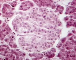
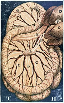
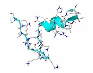
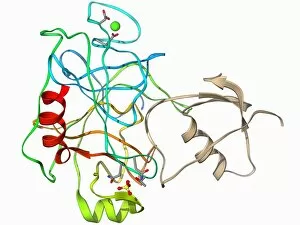
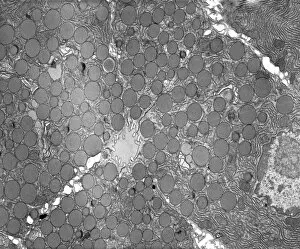
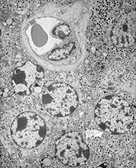
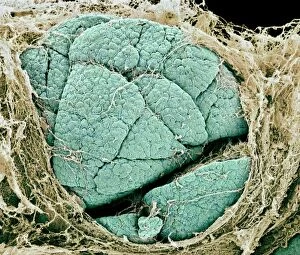
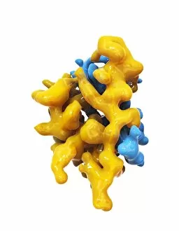
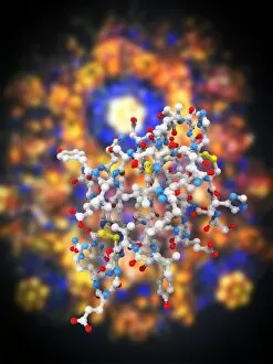
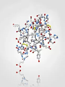
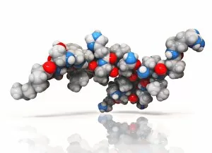
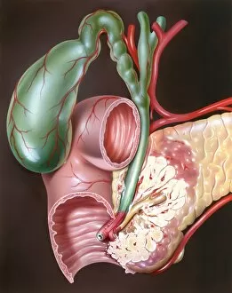
The pancreatic organ, located in the abdomen, is a fascinating and intricate part of our anatomy. Within this complex structure lies the Islet of Langerhans, which plays a crucial role in maintaining our body's balance. In awe-inspiring artwork depicting the pancreas anatomy, we can observe the Islets of Langerhans cells shining like stars amidst a sea of tissue. These microscopic clusters are responsible for producing hormones such as insulin and glucagon that regulate blood sugar levels. Under the lens of a light micrograph, we witness the beauty and intricacy of these Islets of Langerhans. Each cell within them holds immense power to control our metabolism and keep us healthy. A closer look at these incredible cells reveals their unique composition. Ghrelin hormone molecules dance together with insulin molecules C014 / 2121 and F006 / 9761, forming an orchestra that orchestrates our body's energy balance. But it doesn't end there; another molecule called trypsin also takes center stage within the pancreas. Its powerful presence can be seen in stunning artwork alongside its inhibitor counterpart F006 / 9633 - a delicate dance between activation and inhibition to ensure proper digestion. As we marvel at these molecular wonders within the pancreas, let us not forget its overall importance in maintaining our well-being. A color lithograph showcasing "The Pancreas" reminds us how vital this organ is to our survival. So next time you hear about pancreatic health or come across images portraying its complexity, take a moment to appreciate all that goes on behind-the-scenes. The pancreas truly deserves recognition for its remarkable contributions towards keeping us balanced and thriving.