
Exocrine Collection
"Exploring the Intricate World of Exocrine: Unveiling the Pancreas Anatomy through Artwork" Step into a mesmerizing journey through the hidden realms of exocrine


All Professionally Made to Order for Quick Shipping
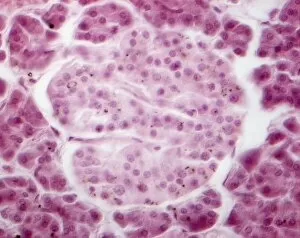
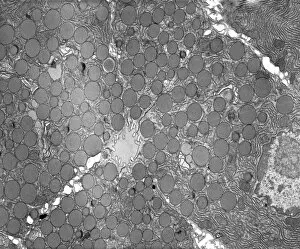
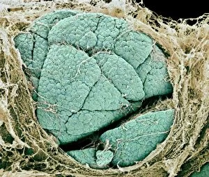
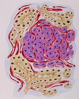
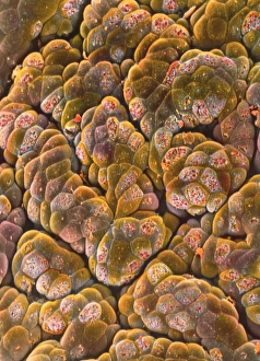
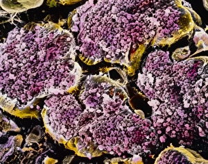
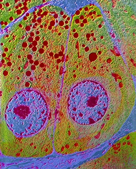
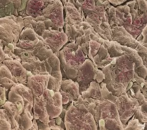
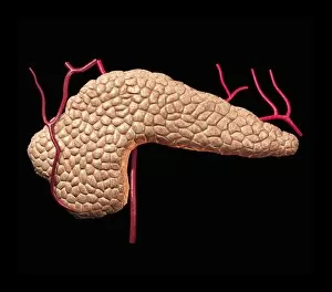
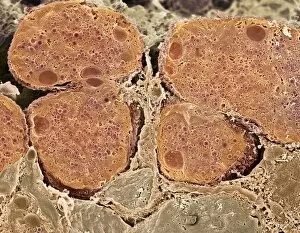
"Exploring the Intricate World of Exocrine: Unveiling the Pancreas Anatomy through Artwork" Step into a mesmerizing journey through the hidden realms of exocrine, as we delve into the intricate anatomy of the pancreas. Through stunning artwork and microscopic imagery, witness the beauty and complexity of this vital organ. The Islet of Langerhans takes center stage in our exploration, captured in breathtaking light micrographs. These tiny clusters of cells hold immense significance in regulating blood sugar levels and insulin production. Marvel at their delicate structure and marvel at their crucial role in maintaining our body's equilibrium. Zooming even closer under a transmission electron microscope (TEM), we uncover the secrets held within pancreatic exocrine cells. The exquisite detail reveals an array of specialized structures that aid in digestion, such as zymogen granules found within pancreatic acinar cells. Witness these remarkable cellular components up close, showcasing nature's ingenuity. Moving beyond microscopy, scanning electron microscope (SEM) images offer a panoramic view of pancreas lobes – like abstract art come to life. Observe their unique shapes and textures as they interconnect to form this essential gland responsible for producing digestive enzymes and hormones. As you immerse yourself in this visual feast, let your imagination wander amidst these captivating visuals that blend science with artistry. Appreciate how each cell contributes to our overall well-being while simultaneously igniting curiosity about our own bodies' inner workings. Exocrine unveils a world often unseen by most but holds profound importance for human health. So take a moment to appreciate its elegance; let it inspire awe for the intricacies woven into every fiber of our being – reminding us just how extraordinary life truly is.