Glass Frame : Leaf midrib, light micrograph


![]()
Mounted Prints from Science Photo Library
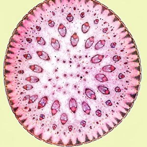
Leaf midrib, light micrograph
Leaf midrib. Light micrograph (LM) of a section through the midrib of a leaf from a monocotyledon plant. The midrib (midvein) is the continuation of a leafs stem along the centre of the leaf. At lower centre is a vascular bundle, which consists of primary xylem (large circles) and primary phloem (green, centre) tissues. Xylem transports water and mineral nutrients from the roots throughout the plant and phloem transports carbohydrate and hormones around the plant. Other, smaller, vascular bundles can also be seen to the left and to the right. The surface (epidermis) of the leaf is covered in a waxy cuticle (red) that helps to prevent water loss
Science Photo Library features Science and Medical images including photos and illustrations
Media ID 6289005
© STEVE GSCHMEISSNER/SCIENCE PHOTO LIBRARY
Axial Cellular Cross Section Cuticle Epidermis False Colour Histological Histology Mid Rib Phloem Spongy Mesophyll Stem Tissue Transverse Vascular Bundle Waxy Xylem Cells False Coloured Light Micrograph Light Microscope Section Sectioned
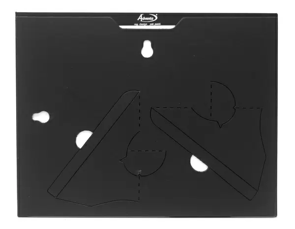
7"x5" Glass Mount
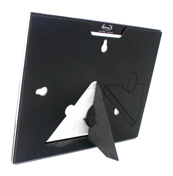
Wall mounted or free-standing, these black edged glass frames feature a smooth chamfered edge and a stylish black border (on back face of the glass). Manufactured from 4mm thick glass, Glass Mounts are a durable, professional way of displaying and protecting your prints. Your 7x5 print is slotted into the back of the frame so can easily be changed if needed.
Tempered Glass Mounts are ideal for wall display, plus the smaller sizes can also be used free-standing via an integral stand
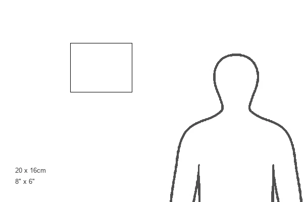
Estimated Image Size (if not cropped) is 17.7cm x 12.7cm (7" x 5")
Estimated Product Size is 20.3cm x 16.2cm (8" x 6.4")
These are individually made so all sizes are approximate
Artwork printed orientated as per the preview above, with landscape (horizontal) orientation to match the source image.
EDITORS COMMENTS
This print showcases the intricate beauty of a leaf midrib, providing us with a fascinating glimpse into the inner workings of a monocotyledon plant. The midrib, also known as the midvein, serves as the central axis along which a leaf's stem extends. In this light micrograph (LM), we can observe various essential components that contribute to the leaf's functionality. At the lower center of the image, we are presented with a prominent vascular bundle composed of primary xylem and primary phloem tissues. These vital structures play distinct roles in transporting water, mineral nutrients, carbohydrates, and hormones throughout the plant. Additional smaller vascular bundles can be observed on both sides. The surface of this leaf is adorned with an enchanting waxy cuticle depicted in red hues. This protective layer acts as a barrier against excessive water loss from evaporation. Through this false-colored LM image captured by Science Photo Library using advanced scanning electron microscopy techniques and histological staining methods, we gain insight into cellular structures such as palisade parenchyma and spongy mesophyll cells within the leaf tissue. Overall, this awe-inspiring photograph not only highlights nature's remarkable complexity but also emphasizes its delicate balance between form and function in sustaining life for these botanical wonders.
MADE IN THE UK
Safe Shipping with 30 Day Money Back Guarantee
FREE PERSONALISATION*
We are proud to offer a range of customisation features including Personalised Captions, Color Filters and Picture Zoom Tools
SECURE PAYMENTS
We happily accept a wide range of payment options so you can pay for the things you need in the way that is most convenient for you
* Options may vary by product and licensing agreement. Zoomed Pictures can be adjusted in the Basket.