Framed Print : Bone growth, light micrograph


![]()
Framed Photos from Science Photo Library
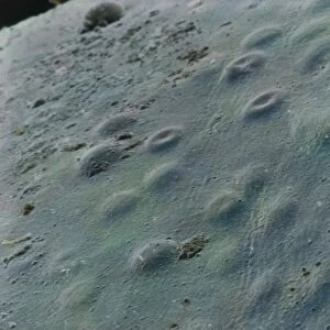
Bone growth, light micrograph
Bone growth. Light micrograph of actively growing cells in the epiphyseal plate (growth plate) between the diaphysis (shaft) and epiphysis (rounded end) of a long bone. Seen at top (pale) is a region of hyaline cartilage cells (chondrocytes) that divide, enlarge and eventually die. The proliferation of these cells is seen at centre (purple). The cartilage matrix is replaced with bone formed from osteoblast cells, which results in a calcified matrix (seen here at bottom, pink). This process is known as ossification and it serves to increase the length of the diaphysis of long bones
Science Photo Library features Science and Medical images including photos and illustrations
Media ID 6419892
© STEVE GSCHMEISSNER/SCIENCE PHOTO LIBRARY
Calcification Cartilage Chondrocyte Chondrocytes Connective Tissue Developing Development Developmental Biology Diaphysis Epiphysis Formation Growing Histology Hyaline Increasing Length Long Matrix Ossification Osteoblast Osteoblasts Osteology Proliferation Shaft Spicule Spicules Spongy Tissue Cells Enlarging Increases Lengthening Light Micrograph Light Microscope Osteogenesis Section Sectioned
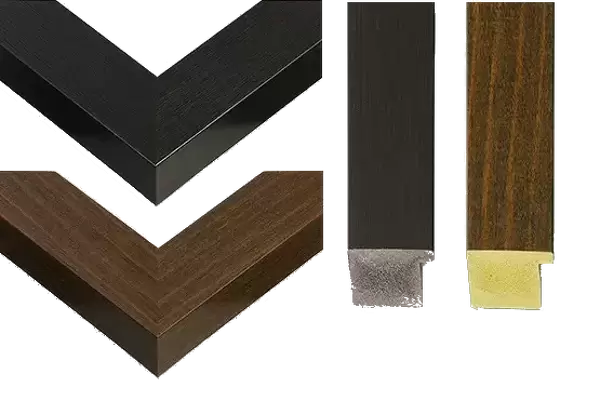
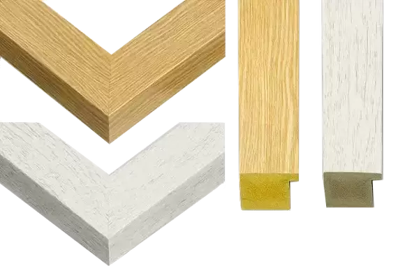
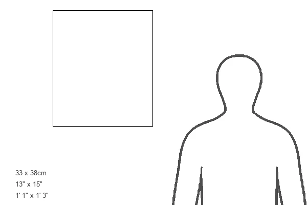
14"x12" (38x32cm) Modern Frame
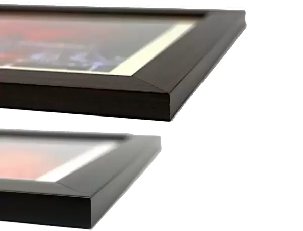
Discover the wonders of science with our Media Storehouse Framed Prints. This captivating piece features a mesmerizing light micrograph image of bone growth, captured by the expert photographers at Science Photo Library. Witness the intricate process of new bone cells forming in the epiphyseal plate, the site of long bone growth. Add this stunning scientific artwork to your home or office to inspire curiosity and ignite conversations. Our high-quality framed prints are meticulously crafted to preserve the vivid colors and details of the original image, ensuring a beautiful addition to any space.
Wood effect frame, card mounted, 10x8 archival quality photo print. Overall outside dimensions 14x12 inches (38x32cm). Environmentally and ozone friendly, 40mm wide x 15mm Polycore® moulding has the look of real wood, is durable and light and easy to hang. Biodegradable and made with non-chlorinated gases (no toxic fumes) it is efficient; producing 100 tons of polystyrene can save 300 tons of trees! Prints are glazed with lightweight, shatterproof, optical clarity acrylic (providing the same general protection from the environment as glass). The back is stapled hardboard with a sawtooth hanger attached. Note: To minimise original artwork cropping, for optimum layout, and to ensure print is secure, the visible print may be marginally smaller
Contemporary Framed and Mounted Prints - Professionally Made and Ready to Hang
Estimated Image Size (if not cropped) is 17.1cm x 24.4cm (6.7" x 9.6")
Estimated Product Size is 32.5cm x 37.6cm (12.8" x 14.8")
These are individually made so all sizes are approximate
Artwork printed orientated as per the preview above, with portrait (vertical) orientation to match the source image.
EDITORS COMMENTS
This print showcases the intricate process of bone growth at a microscopic level. The light micrograph reveals the dynamic activity occurring in the epiphyseal plate, which is located between the diaphysis and epiphysis of a long bone. At the top, we observe a region filled with pale hyaline cartilage cells known as chondrocytes. These cells divide, enlarge, and eventually perish as part of their natural life cycle. In the center of this image, we witness an awe-inspiring proliferation of these growing cells in a striking purple hue. This vibrant display represents the active formation and development taking place within this vital tissue. As time progresses, osteoblast cells step forward to replace the cartilage matrix with solid bone material. At the bottom section of this micrograph lies an eye-catching pink calcified matrix that signifies successful ossification – a crucial process responsible for lengthening our long bones throughout development. By depositing new layers upon layers of bone material, our bodies ensure continuous growth and maintain healthy anatomical structures. This remarkable visual insight into bone growth offers us valuable knowledge about cellular dynamics and highlights how our bodies naturally adapt to increase both strength and stature over time. It serves as a testament to nature's incredible ability to construct complex tissues from simple building blocks while maintaining harmony within our biological systems.
MADE IN THE UK
Safe Shipping with 30 Day Money Back Guarantee
FREE PERSONALISATION*
We are proud to offer a range of customisation features including Personalised Captions, Color Filters and Picture Zoom Tools
SECURE PAYMENTS
We happily accept a wide range of payment options so you can pay for the things you need in the way that is most convenient for you
* Options may vary by product and licensing agreement. Zoomed Pictures can be adjusted in the Basket.