
Cartilage Collection
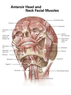
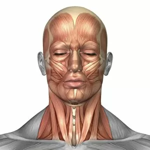
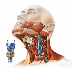
"Exploring the Foundation of Flexibility: The Marvels of Cartilage" Facial muscles of the human face (with labels


All Professionally Made to Order for Quick Shipping
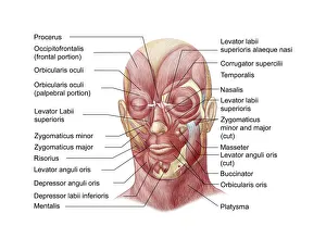
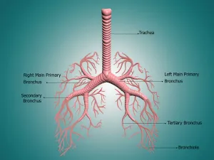
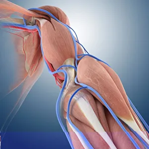
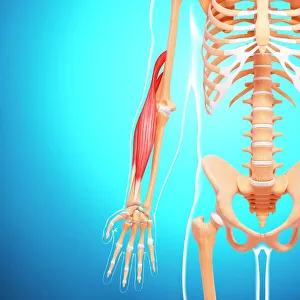
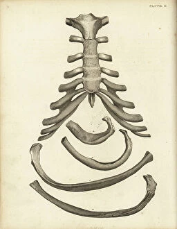
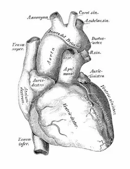
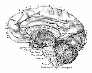
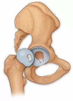
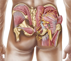
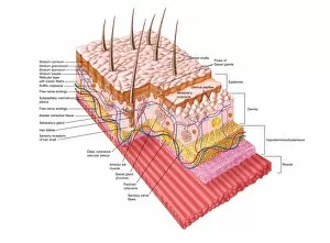
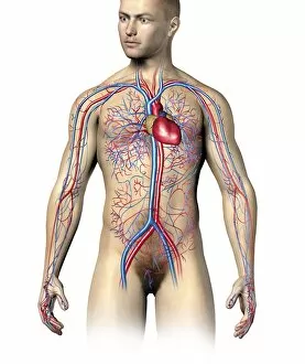
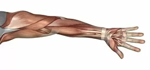
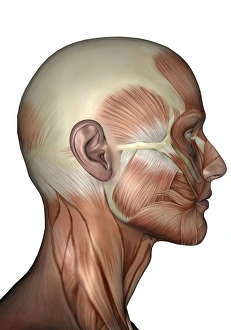
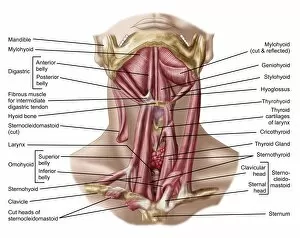
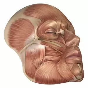
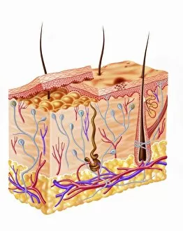
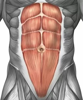
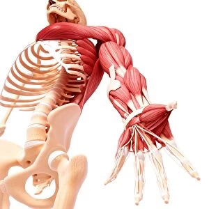
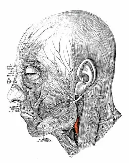
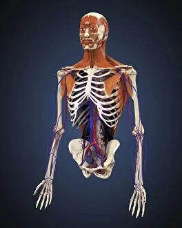
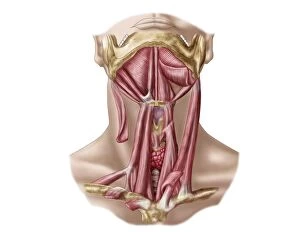
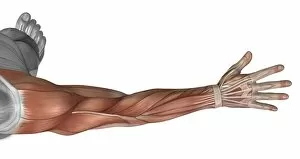
"Exploring the Foundation of Flexibility: The Marvels of Cartilage" Facial muscles of the human face (with labels): Delve into the intricate network that supports our facial expressions, allowing us to smile, frown, and express a myriad of emotions. Anatomy of human knee joint: Witness the resilience and shock-absorbing capabilities in our knees as it cushions each step we take, ensuring smooth movement and preventing bone-on-bone friction. The human body with superimposed colored plates by Julien Bougle: Uncover the hidden beauty within our bodies as vibrant hues highlight the vital role played by cartilage in maintaining structural integrity and flexibility. Anatomy of the bronchus and bronchial tubes: Discover how cartilaginous rings keep our airways open like sturdy scaffolding, facilitating effortless breathing while protecting against collapse. Arm circulation, anatomical artwork C013 / 7419: Explore how blood vessels intertwine with resilient cartilage structures in our arms, nourishing every muscle fiber to sustain their strength and mobility. Muscles of the neck: Peer beneath the surface to witness how cartilaginous discs provide stability between vertebrae in our necks, enabling fluid movements while safeguarding delicate nerves. Human arm musculature, artwork F007 / 1810: Marvel at how interconnected layers of muscles rely on supportive cartilage for optimal function—allowing us to lift weights or embrace loved ones with ease. Slipped disc: Understand why proper care is essential as we uncover what happens when intervertebral discs made up partly by fibrocartilage shift out-of-place—causing discomfort but also highlighting its importance for spinal health. Human anatomy scientific illustrations: heart, veins and arteries: Journey through an intricately illustrated world where blood vessels intertwine alongside cardiac valves supported by cartilaginous structures, ensuring efficient circulation.