
Diaphysis Collection
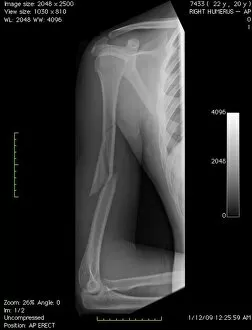
"Exploring the Diaphysis: Unveiling the Inner Structure of Long Bones" A broken arm bone reveals the intricate diaphysis, or shaft


All Professionally Made to Order for Quick Shipping
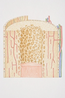
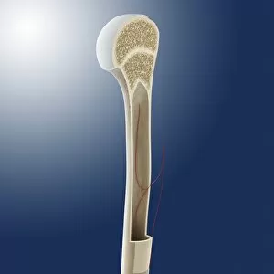
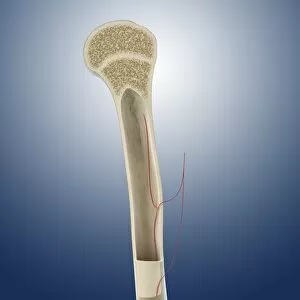
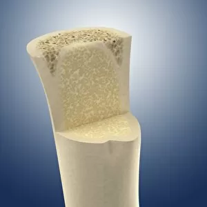
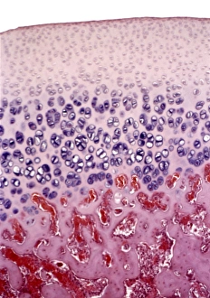
"Exploring the Diaphysis: Unveiling the Inner Structure of Long Bones" A broken arm bone reveals the intricate diaphysis, or shaft, that provides strength and support to our limbs. With a digital X-ray, we can now visualize the cross-section diagram of a human long bone, highlighting its diaphysis and surrounding structures. Delving into early life, a biomedical illustration showcases the cross section of a newborn baby's long bone diaphysis during development. Witnessing childhood growth, another biomedical illustration displays the cross section of a developing long bone's diaphysis as it matures over time. Finally reaching adulthood, an insightful biomedical illustration unveils the fully developed long bone's diaphysis in all its complexity and strength. Journeying deeper within bones, explore the fascinating anatomy of human bone marrow residing within these remarkable structures. Marvel at artwork depicting Paprosky femur defect type IIIA lateral view - showcasing how even complex conditions affect this crucial part of our bones' structure. Intriguingly detailed artwork C016 / 2888 unravels the secrets hidden within humeral bone marrow cavity - shedding light on its vital role in our skeletal system. Continuing exploration with artwork C016 / 2887 revealing further insights into humeral bone marrow cavity - unlocking knowledge about its function and composition. Peering inside through artwork C016 / 2886 captures an extraordinary glimpse into the interior of long bones - where essential processes occur for our overall health and mobility. Witnessing growth at a microscopic level, delve into mesmerizing light micrographs capturing intricate details of bone growth within their diaphyses.