
Calcification Collection
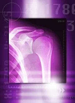
"Exploring the Intricate World of Calcification: From Ancient Engravings to Modern Medical Imaging" Tendinitis of the shoulder


All Professionally Made to Order for Quick Shipping
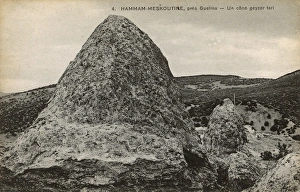
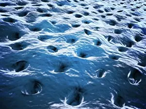
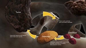
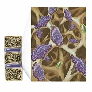
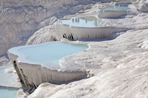
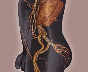
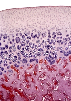
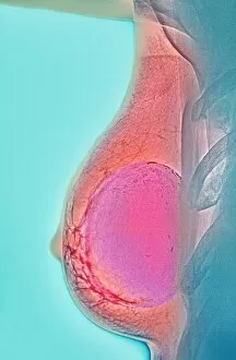
"Exploring the Intricate World of Calcification: From Ancient Engravings to Modern Medical Imaging" Tendinitis of the shoulder? X-ray reveals calcification as a possible cause for your discomfort. Did you know kidney stones have been troubling humans since the 18th century? Calcification plays a major role in their formation. Unveiling mysteries with colored MRI brain scans: Sturge-Weber syndrome showcases abnormal calcifications within the brain. Journey back in time through an engraving titled "Four pour la calcination des os. " Discover how ancient civilizations understood bone calcification. Witness nature's artistry at Hammam Maskhoutine, Algeria, where a dried geyser cone stands as a testament to mineral-rich waters and natural calcification processes. Aortic dissection captured in stunning detail through a 3D CT scan, highlighting potential complications caused by arterial wall calcifications. Pamukkale Geothermal Area mesmerizes with its surreal white terraces formed by calcium-rich hot springs – an exquisite example of geological calcification wonders. Osteoclasts tirelessly break down bone tissue during remodeling, ensuring healthy skeletal maintenance throughout our lives – microscopic view unveils their work. Delving deeper into dental health: behold the intricate beauty of dentine under the microscope, showcasing natural tooth structure and its unique patterns of calcification. Balancing act between osteoclasts and osteoblasts. Witness these remarkable cells collaborating to build healthy bones while maintaining optimal levels of calcium deposition and resorption. Bone metastasis demystified. Conceptual image illustrates how cancer cells exploit normal bone remodeling processes for their own growth – shedding light on this complex form of pathological calcification.