Home > Science > Xray
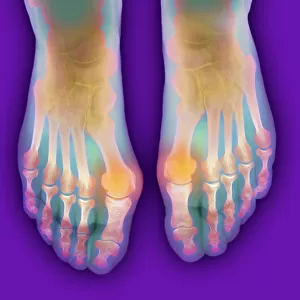
Hallux rigidus before surgery, X-ray

![]()
Wall Art and Photo Gifts from Science Photo Library
Hallux rigidus before surgery, X-ray
Hallux rigidus before surgery. X-ray showing foot bones before surgery to correct a case of hallux rigidus, a stiff big toe. This is due to a bone spur on the metatarsophalangeal (MTP) joint of the toe, but can also be due to arthritis. The surgery carried out was a right dorsal cheilectomy (to remove the bony lump) and a hemi-arthroplasty (repairing or replacing half the joint). The lump on the big toe can be seen as a bony protrusion (white) at upper left, at the joint where the 1st metatarsal and proximal phalanx meet. For the operation, see images C014/7778 to C014/7780
Science Photo Library features Science and Medical images including photos and illustrations
Media ID 9223147
© ANTONIA REEVE/SCIENCE PHOTO LIBRARY
Big Toe Deformed Deformity Diagnosing Diagnosis Diagnostic Diagnostics Foot From Above Joint Metatarsal Metatarsals Orthopaedics Patient Phalanges Radiography Superior Toes X Ray Machine Xray Right Foot
EDITORS COMMENTS
This print from Science Photo Library showcases a pre-surgery X-ray of hallux rigidus, a condition characterized by a stiff big toe. The image reveals the foot bones before undergoing corrective surgery to address this deformity caused by either arthritis or a bone spur on the metatarsophalangeal (MTP) joint of the toe. The surgical procedure performed was a right dorsal cheilectomy, involving the removal of the bony lump, and a hemi-arthroplasty, which repaired or replaced half of the affected joint. In this monochrome X-ray, we can clearly observe the bony protrusion at the upper left corner where the first metatarsal and proximal phalanx meet. With superior diagnostic capabilities provided by radiography and an advanced x-ray machine, orthopedic specialists were able to accurately diagnose and assess this patient's condition. This image serves as both an educational tool for medical professionals in understanding hallux rigidus and as evidence of successful treatment options available. Antonia Reeve skillfully captures not only the clinical aspects but also conveys compassion towards patients suffering from such conditions. It is important to note that this caption does not promote any commercial use nor mention any specific company; instead, it focuses on highlighting key medical terms associated with hallux rigidus diagnosis, treatment procedures, and anatomical structures involved in this particular case.
MADE IN THE UK
Safe Shipping with 30 Day Money Back Guarantee
FREE PERSONALISATION*
We are proud to offer a range of customisation features including Personalised Captions, Color Filters and Picture Zoom Tools
SECURE PAYMENTS
We happily accept a wide range of payment options so you can pay for the things you need in the way that is most convenient for you
* Options may vary by product and licensing agreement. Zoomed Pictures can be adjusted in the Basket.