Fraud Blocker Skip to main content

Diagnostic Collection

"Unlocking the Secrets of Health: Exploring the World Tools" Medieval Urine Wheel: From ancient times to modern medicine

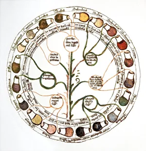
Background imageDiagnostic Collection: Medieval urine wheel

Medieval urine wheel. Labelled in Latin, this 15th century diagram shows some of the possible colours of urine (outer edge of circle) to help doctors diagnose urine-related diseases

Background imageDiagnostic Collection: Eyesight test chart

Eyesight test chart

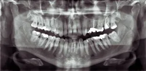
Background imageDiagnostic Collection: Panoramic dental X-ray

Panoramic dental X-ray showing all the teeth in the upper and lower jaws

Background imageDiagnostic Collection: Broken wrist bone, X-ray C017 / 7187

Broken wrist bone, X-ray C017 / 7187
Broken wrist bone. X-ray of the hand of a 12 year old male with a fractured trapezium bone. The trapezium is the leftmost bone in the group of bones at the base of the bad

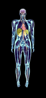
Background imageDiagnostic Collection: Full body scan, MRI scan

Full body scan, MRI scan
Full body scan of a female swimmer. Her brain is purple (top), her heart and arteries are red/orange (upper centre), her kidneys are pale green (centre)

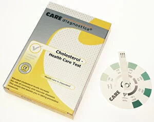
Background imageDiagnostic Collection: Home cholesterol test kit

Home cholesterol test kit
Cholesterol indicator test for use at home. This kit tests a small sample of blood for the presence of high levels of cholesterol. The scale on the disk indicates the level of cholesterol

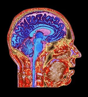
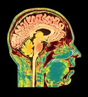
Background imageDiagnostic Collection: Brain anatomy, MRI scan

Brain anatomy, MRI scan
Brain anatomy. Coloured magnetic resonance imaging (MRI) scan of the human head from the side. The sagittal scan has halved the brain, revealing the internal anatomy

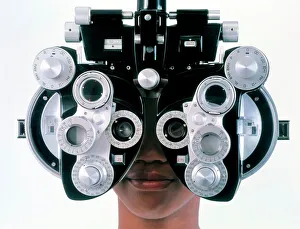
Background imageDiagnostic Collection: Eye examination

Eye examination
MODEL RELEASED. MODEL RELEASED. Eye examination. Teenage (16 year old) girl looks through a refractor during an eye examination at an ophthalmologist

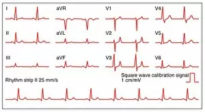
Background imageDiagnostic Collection: ECGs of a normal heart rate, artwork

ECGs of a normal heart rate, artwork
Electrocardiograms (ECG) of a normal heart rate, artwork. An ECG measures the electrical activity of the heart. The wave on the ECG appears upwards or downwards depending upon whether the electrical

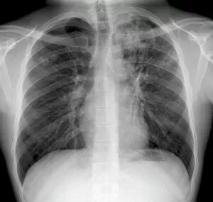
Background imageDiagnostic Collection: Tuberculosis, X-ray

Tuberculosis, X-ray
Tuberculosis. X-ray of the chest of a 25 year old male patient with pulmonary tuberculosis. Affected areas of the lungs (dark areas) are shown by grainy white patches

Background imageDiagnostic Collection: Brain tumour, MRI scan

Brain tumour, MRI scan
Brain tumour. Coloured orthogonal views (axial left, coronal middle, sagittal right) of a male head, made from magnetic resonance imaging (MRI) scans

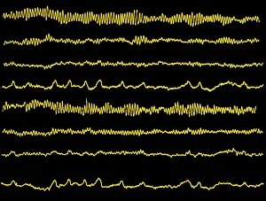
Background imageDiagnostic Collection: Normal EEG read out of the brains alpha waves

Normal EEG read out of the brains alpha waves
Normal electroencephalogram (EEG) read-out of the brains alpha waves. original is b/w m400/01m

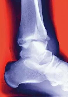
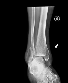
Background imageDiagnostic Collection: Fractured ankle, X-ray

Fractured ankle, X-ray
Fractured ankle. Coloured profile X-ray of a distal fibula fracture (upper centre). The fibula is the smaller leg bone running down from top centre. The larger bone is the tibia

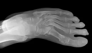
Background imageDiagnostic Collection: Lisfranc fracture, X-ray

Lisfranc fracture, X-ray

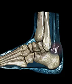
Background imageDiagnostic Collection: Ruptured Achilles tendon, MRI C018 / 0649

Ruptured Achilles tendon, MRI C018 / 0649
Ruptured Achilles tendon. Coloured magnetic resonance imaging (MRI) scan of the foot of a 25 year old patient with a ruptured Achilles tendon

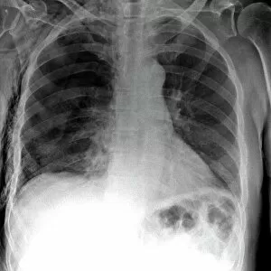
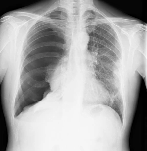
Background imageDiagnostic Collection: Tension pneumothorax, X-ray

Tension pneumothorax, X-ray
Tension pneumothorax. X-ray of the chest of a patient with tension pneumothorax that has lead to soft-tissue emphysema. Tension pneumothorax is a progressive deterioration

Background imageDiagnostic Collection: Normal human brain, MRI scan C016 / 8845

Normal human brain, MRI scan C016 / 8845
Brain. Coloured magnetic resonance imaging (MRI) scan of a sagittal section through a patients head showing a healthy human brain

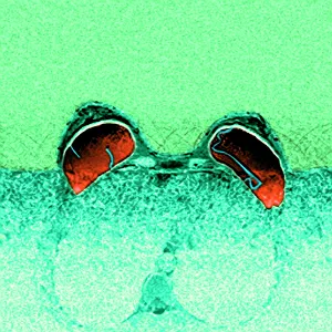
Background imageDiagnostic Collection: Ruptured breast implants, MRI

Ruptured breast implants, MRI
Ruptured breast implants. Coloured magnetic resonance imaging (MRI) scan of ruptures in both of a womans breast implants (red). These are intracapsular ruptures, the most common type

Background imageDiagnostic Collection: Phrenology bust

Phrenology bust. Phrenology was a popular pseudoscience of the nineteenth century. The basis for the theory lay in a misplaced belief that a persons character

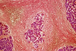
Background imageDiagnostic Collection: Liver tissue cirrhosis, light micrograph

Liver tissue cirrhosis, light micrograph
Liver cirrhosis. Light micrograph of a section through liver tissue showing cirrhosis. This is a condition where the liver responds to the injury or death of some of its cells by producing

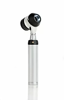
Background imageDiagnostic Collection: Dermatoscope

Dermatoscope. This instrument is used to examine the skin. It includes a magnifier and a light source

Background imageDiagnostic Collection: Normal human brain, MRI scan C016 / 8850

Normal human brain, MRI scan C016 / 8850
Brain. Coloured magnetic resonance imaging (MRI) scan of a sagittal section through a patients head showing a healthy human brain

Background imageDiagnostic Collection: Neck, side view, coloured X-ray

Neck, side view, coloured X-ray
Neck from the side, coloured X-ray. The front of the body is at right. The spine runs down centre. It is made up of block-like bones called vertebrae

Background imageDiagnostic Collection: Healthy heart, conceptual image

Healthy heart, conceptual image
MODEL RELEASED. Healthy heart. Conceptual image of a healthy heart diagnosis represented by a doctor giving a patient a heart shape

Background imageDiagnostic Collection: Slipped disc

Slipped disc. Spinal model demonstrating a slipped intervertebral disc. The slipped disc is the small bulge (red, centre), near the base of the spine

Background imageDiagnostic Collection: Lung function test

Lung function test. Spirometer sitting on lung function test results

Background imageDiagnostic Collection: Inflamed spinal discs, MRI scan

Inflamed spinal discs, MRI scan
Inflamed spinal discs. Coloured magnetic resonance imaging (MRI) scan of a side view of the spine of a patient with discitis. The front of the body is at left, the skull is at top

Background imageDiagnostic Collection: Robert Koch (1843-1910) German bacteriologist and physician in his laboratory

Robert Koch (1843-1910) German bacteriologist and physician in his laboratory
530414 Robert Koch (1843-1910) German bacteriologist and physician in his laboratory. In 1890 Koch introduced Tuberculin which he thought was a cure for Tuberculosis
Background imageDiagnostic Collection: Broken ankle, X-ray C017 / 7185

Broken ankle, X-ray C017 / 7185
Broken ankle. X-ray of the lower leg of an 85 year old female with fractures at the ends of the tibia (shin bone, right) and fibula (calf bone, left)

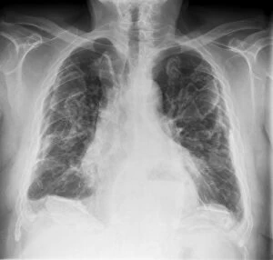
Background imageDiagnostic Collection: Asbestosis, X-ray C017 / 7143

Asbestosis, X-ray C017 / 7143
Asbestosis. X-ray of the chest of a 78 year old male that has been exposed to asbestos, showing diffuse plaques (light grey areas) in the lungs (dark)

Background imageDiagnostic Collection: Pneumothorax, X-ray C017 / 7148

Pneumothorax, X-ray C017 / 7148
Pneumothorax. X-ray of the chest of a 50 year old male patient with a pneumothorax, or collapsed lung. The right lung (left on X-ray) has collapsed due to a build-up of air (solid grey)

Background imageDiagnostic Collection: Arthritis of the neck, X-ray C017 / 7389

Arthritis of the neck, X-ray C017 / 7389
Arthritis of the neck. X-ray of the neck of a 67 year old female with osteoarthritis of the neck. Osteoarthritis of the neck, also known as cervical spondylosis

Background imageDiagnostic Collection: Stroke, MRI and 3D CT scans C016 / 6419

Stroke, MRI and 3D CT scans C016 / 6419
Stroke. Coloured composite image of a magnetic resonance imaging (MRI) scan of the brain and 2D and 3D computed tomography (CT)

Background imageDiagnostic Collection: Normal human brain, MRI scan C016 / 8843

Normal human brain, MRI scan C016 / 8843
Brain. Coloured magnetic resonance imaging (MRI) scan of a sagittal section through a healthy human brain. The highly folded area (brown) is the cerebrum, which is responsible for conscious thought

Background imageDiagnostic Collection: Normal skull and brain, 3D CT scan C016 / 6333

Normal skull and brain, 3D CT scan C016 / 6333
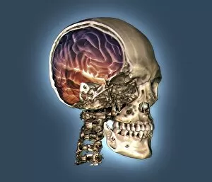
Normal skull and brain. Coloured 3D computed tomography (CT) scan of the head of a 35 year old patient. Part of the skull has been cut-away to show the brain (purple and red)

Background imageDiagnostic Collection: Normal head and neck, MRI and 3D CT scans C016 / 6337

Normal head and neck, MRI and 3D CT scans C016 / 6337
Normal head. Coloured composite image of a magnetic resonance imaging (MRI) scan of the brain and a 3D computed tomography (CT) scan of the head and neck of a 35 year old patient

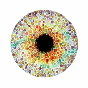
Background imageDiagnostic Collection: Colour blindness, conceptual image

Colour blindness, conceptual image. Composite image of a human eye and an Ishihara colour test card used to check for red-green colour blindness

Background imageDiagnostic Collection: Broken leg, X-ray C017 / 7977

Broken leg, X-ray C017 / 7977
Broken leg. X-ray of the leg of a 32 year old male who has been in a parachuting accident. The fibula (calf bone, right) is fractured

Background imageDiagnostic Collection: Broken foot, X-ray C017 / 7975

Broken foot, X-ray C017 / 7975
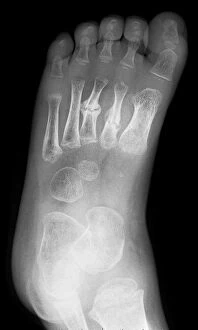
Broken foot. X-ray of the foot of a 3 year old male patient with fractured second and third metatarsals (foot bones)

Background imageDiagnostic Collection: Pinned broken arm, X-ray C017 / 7869

Pinned broken arm, X-ray C017 / 7869
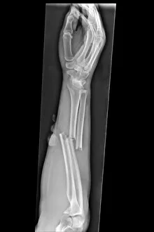
Pinned broken arm. X-ray of the arm of a patient that has been internally fixed after fracturing both of the lower arm bones, the ulna and radius

Background imageDiagnostic Collection: Broken arm, X-ray C017 / 7863

Broken arm, X-ray C017 / 7863
Broken arm. X-ray of the arm of a patient that has broken both of the lower arm bones, the ulna and radius

Background imageDiagnostic Collection: Liver mass, ultrasound scan C017 / 7780

Liver mass, ultrasound scan C017 / 7780
Liver mass. Ultrasound scan of the liver of a 69 year old male patient showing an irregular mass (centre)

Background imageDiagnostic Collection: Secondary liver cancer, ultrasound scan C017 / 7764

Secondary liver cancer, ultrasound scan C017 / 7764
Secondary liver cancer. Ultrasound scan of a 63 year old male patients liver showing a metastatic (secondary) tumour. The cancer has metastasised (spread) from a primary cancer

Background imageDiagnostic Collection: Dental plaque and tartar

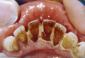
Dental plaque and tartar. Clinical view of a the teeth in a patients lower jaw, with the incisors coated in bacterial plaque and tartar (brown)

Background imageDiagnostic Collection: Caricature of Roentgen and X-rays

Caricature of Roentgen and X-rays. The German experimental physicist Wilhelm Konrad Roentgen (1845-1923) had discovered X-rays in 1895. This artwork shows his skeleton revealed by the use of X-rays

Background imageDiagnostic Collection: Broken arm, X-ray

Broken arm, X-ray
Broken arm. X-ray of the forearm of a 29 year old male patient with a fractured ulna. Treatment is with immobilisation of the bone, either externally using a cast, or internally using pins

Background imageDiagnostic Collection: Fractured vertebra, X-ray C017 / 7586

Fractured vertebra, X-ray C017 / 7586
Fractured vertebra. X-ray of the lower spine of a 50 year old male with a fracture to the L1 lumbar vertebra (thord block from top)

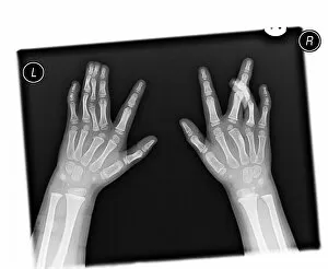
Background imageDiagnostic Collection: Deformed hands, X-ray C017 / 7561

Deformed hands, X-ray C017 / 7561
Deformed hands. X-ray of the hands of a 7 year old female patient with congenital (present at birth) abnormalities of the fingers


All Professionally Made to Order for Quick Shipping

"Unlocking the Secrets of Health: Exploring the World Tools" Medieval Urine Wheel: From ancient times to modern medicine, urine analysis has been a diagnostic tool used to detect various health conditions. Eyesight Test Chart: A simple yet crucial diagnostic tool that helps determine visual acuity and identify potential eye problems. Broken Wrist Bone, X-ray C017 / 7187: X-rays provide a clear view of fractured bones, aiding in accurate diagnosis and guiding treatment plans. Panoramic Dental X-ray: This comprehensive imaging technique reveals dental issues like cavities, impacted teeth, or jaw abnormalities for effective treatment planning. Home Cholesterol Test Kit: Empowering individuals with easy-to-use kits to monitor cholesterol levels at home and take proactive steps towards heart health. Full Body Scan, MRI Scan: Non-invasive and highly detailed imaging technology that allows doctors to visualize internal organs and diagnose complex medical conditions accurately. Brain Anatomy, MRI Scan: Advanced brain imaging enables neurologists to study intricate structures within the brain for precise diagnoses of neurological disorders. ECGs of a Normal Heart Rate, Artwork: Electrocardiograms help assess heart function by recording electrical activity; this artwork showcases what a healthy heartbeat looks like on paper. Tuberculosis, X-ray: Chest x-rays play a vital role in diagnosing tuberculosis by detecting characteristic lung abnormalities associated with this infectious disease. Normal EEG Readout of the Brain's Alpha Waves: Electroencephalography records brain wave patterns helping neurologists evaluate brain function and diagnose epilepsy or sleep disorders effectively. Eye Examination:A thorough examination assessing vision sharpness (visual acuity), peripheral vision tests (visual field), color perception evaluation among others is essential for maintaining good eye health Fractured Ankle, X-Ray.