Fraud Blocker Skip to main content

Phalanges Collection

"Unveiling the Marvels of Phalanges: A Journey into the Intricacies of Human Anatomy" Step into the world of phalanges

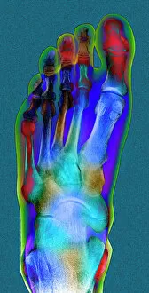
Background imagePhalanges Collection: Normal foot, X-ray

Normal foot, X-ray
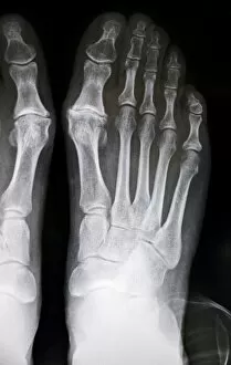
Normal foot. Coloured X-ray of a healthy human foot seen from the top (antero-posterior or dorso- plantar projection). The foot comprises 26 bones and two sesamoid bones (not seen)

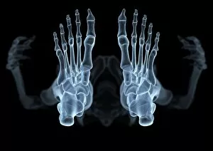
Background imagePhalanges Collection: Skeleton from below, X-ray artwork

Skeleton from below, X-ray artwork
Skeleton. Computer x-ray artwork of a human skeleton viewed from below

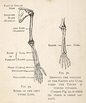
Background imagePhalanges Collection: Diagrams of the bones of hand and arm

Diagrams of the bones of hand and arm
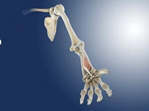
Diagrams of the bones of the left arm and hand, showing the position of the radius and ulna when the thumb is turned inwards. The shoulder blade and part of the collar bone can also be seen

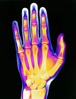
Background imagePhalanges Collection: Coloured X-ray of the healthy hand of a man

Coloured X-ray of the healthy hand of a man
Mans hand. Coloured X-ray of the healthy hand of a man, showing the arrangement of its bones. An outline of the flesh of the hand is seen

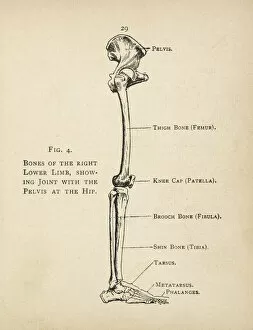
Background imagePhalanges Collection: Diagram of the bones of the right leg and hip

Diagram of the bones of the right leg and hip
Diagram of the bones of the right leg, showing the joint with the pelvis at the hip

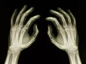
Background imagePhalanges Collection: X-ray of healthy human hands

X-ray of healthy human hands
Healthy hands. X-ray of healthy human hands. The bones are white and the tissue is pale grey. There are two phalanges bones in each of the thumbs and three in each finger

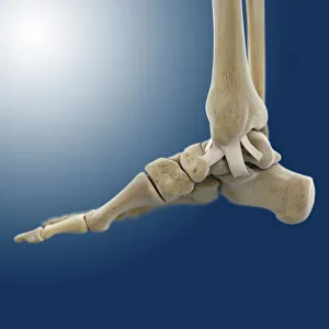
Background imagePhalanges Collection: Outer ankle ligaments, artwork C013 / 4452

Outer ankle ligaments, artwork C013 / 4452
Outer ankle ligaments. Computer artwork of the bones and ligaments (white) of the right foot and ankle seen from the side

Background imagePhalanges Collection: Australopithecine or Homo habilis foot (OH8) cast

Australopithecine or Homo habilis foot (OH8) cast
Cast of a near complete foot (OH 8) from an Australopthecus or Homo habilis discovered at Olduvai Gorge, Tanzania by Louis Leakey in 1960. It dates back to 1.75 million years ago

Background imagePhalanges Collection: Inner ankle ligaments, artwork C013 / 4451

Inner ankle ligaments, artwork C013 / 4451
Inner ankle ligaments. Computer artwork of the bones and ligaments (white) of the right foot and ankle seen from the side

Background imagePhalanges Collection: Comparing the bones in the hands of nine different mammals, 1898 (colour litho)

Comparing the bones in the hands of nine different mammals, 1898 (colour litho)
1065622 Comparing the bones in the hands of nine different mammals, 1898 (colour litho) by Haeckel, Ernst (1834-1919); Private Collection; (add.info.: 1.Man, 2.Gorilla, 3. Orangutang, 4. dog, 5)

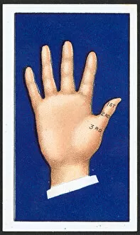
Background imagePhalanges Collection: Thumb (colour litho)

Thumb (colour litho)
2800169 Thumb (colour litho) by English School, (20th century); Private Collection; (add.info.: Thumb. 1st, 2nd and 3rd phalanges)

Background imagePhalanges Collection: Pairs of phalanges (bronze)

Pairs of phalanges (bronze)
614971 Pairs of phalanges (bronze) by Roman; Museo Archeologico Nazionale, Florence, Italy; (add.info.: Coppia di falangi); Photo eNicol Orsi Battaglini; out of copyright

Background imagePhalanges Collection: Hand muscle anatomy, 1831 artwork

Hand muscle anatomy, 1831 artwork
Hand muscle anatomy. Radial view of the ligaments and muscles of the hand (main artwork, centre). The insets show various aspects of the ligaments, muscles, and bones of the fingers

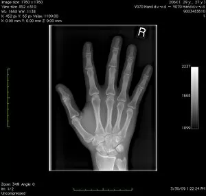
Background imagePhalanges Collection: Normal hand, digital X-ray

Normal hand, digital X-ray
Normal hand. Digital X-ray of the normal right hand of a patient in their late twenties. Various sets of data relating to the image can be seen around the edges of the X-ray

Background imagePhalanges Collection: Amputated toes, X-ray C017 / 7166

Amputated toes, X-ray C017 / 7166
Amputated toe. X-ray of the left foot of a 75 year old male patient where the tip of the big toe and two thirds of the next toe have been amputated

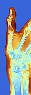
Background imagePhalanges Collection: Arthrosis of the hand, X-ray F006 / 4616

Arthrosis of the hand, X-ray F006 / 4616
Coloured X-ray of the hand of a patient with arthrosis, particularly the saddle joint. Arthrosis is a degenerative joint disease that damages cartilage and bone

Background imagePhalanges Collection: Amputated fingers, X-ray C017 / 7150

Amputated fingers, X-ray C017 / 7150
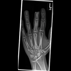
Amputated fingers. X-ray of the right hand of a 57 year old male patient where the tips of the fourth and fifth fingers have been amputated

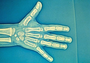
Background imagePhalanges Collection: Child hand X-ray

Child hand X-ray
Childs hand bones (3 year old). Coloured X-ray showing the bones in the hand of a three year old child. Bones appear white

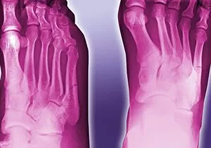
Background imagePhalanges Collection: Fractured foot, coloured X-ray

Fractured foot, coloured X-ray
Fractured foot. Coloured X-rays of a fractured fifth metatarsal (to the right of each image). The fracture is seen as a small black crack

Background imagePhalanges Collection: Childs foot, X-ray

Childs foot, X-ray
Childs foot. Coloured X-ray of the healthy right foot of an eight year old boy. The toes are made up of the phalanx bones (phalanges), two for the big toe (lower right) and three for the others

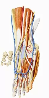
Background imagePhalanges Collection: Foot anatomy

Foot anatomy, computer artwork. Muscles are pink, tendons and ligaments are white and bones are cream. Tendons and ligaments are bands of inelastic fibrous tissue

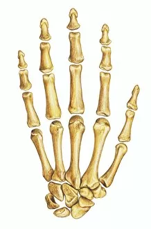
Background imagePhalanges Collection: Illustration of right hand bones

Illustration of right hand bones
Medicine: Human anatomy, right hand bones. Drawing

Background imagePhalanges Collection: Illustration of hand and foot bones

Illustration of hand and foot bones
Medicine: Human anatomy, hand and foot bones

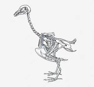
Background imagePhalanges Collection: anatomy, animal themes, bird, coracoid, cranium, cut out, femur, humerous, mandible

anatomy, animal themes, bird, coracoid, cranium, cut out, femur, humerous, mandible, no people, ostrich, pelvis girdle, pen and ink, phalanges, physiology, profile, pygostyle, side view, skeleton

Background imagePhalanges Collection: Human foot, internal anatomy

Human foot, internal anatomy

Background imagePhalanges Collection: Cross section biomedical illustration of fingernail

Cross section biomedical illustration of fingernail

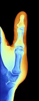
Background imagePhalanges Collection: Arthrosis of the hand, X-ray F006 / 4605

Arthrosis of the hand, X-ray F006 / 4605
Coloured X-ray of the hand of a patient with arthrosis, particularly the saddle joint. Arthrosis is a degenerative joint disease that damages cartilage and bone

Background imagePhalanges Collection: Arthrosis of the hand, X-ray F006 / 4595

Arthrosis of the hand, X-ray F006 / 4595
Coloured X-ray of the hand of a patient with arthrosis of the hands. particularly the saddle joint. Arthrosis is a degenerative joint disease that damages cartilage and bone

Background imagePhalanges Collection: Arthrosis of the hand, X-ray F006 / 4598

Arthrosis of the hand, X-ray F006 / 4598
Coloured X-ray of the hand of a patient with arthrosis, particularly the saddle joint. Arthrosis is a degenerative joint disease that damages cartilage and bone

Background imagePhalanges Collection: Dislocated finger, X-ray C017 / 7183

Dislocated finger, X-ray C017 / 7183
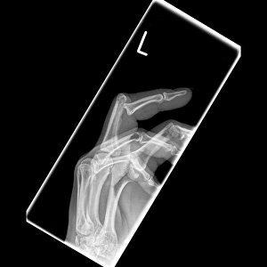
Dislocated finger. X-ray of the left hand of a 51 year old female patient with a dislocated middle finger. Once the bones have been manipulated back into place treatment is with immobilisation

Background imagePhalanges Collection: Foot ligaments and tendons, artwork C016 / 7010

Foot ligaments and tendons, artwork C016 / 7010
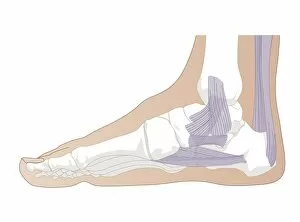
Foot ligaments and tendons. Artwork of the bones, ligaments (white, between bones), and tendons (white, muscle-bone) of the foot and the ankle joint from the side

Background imagePhalanges Collection: Hand ligaments and tendons, artwork

Hand ligaments and tendons, artwork
Hand bones and cartilage. Artwork of the bones and cartilage (white, ends of bones) of the hand and the wrist joint from the rear (dorsal) side

Background imagePhalanges Collection: Hand anatomy, illustration C018 / 0796

Hand anatomy, illustration C018 / 0796
Hand anatomy. Illustration showing the bones, nerves and blood vessels of the hand. There are 27 bones, made up of 14 phalanges on the fingers and thumb

Background imagePhalanges Collection: Radiologist examining a hand X-ray C014 / 1289

Radiologist examining a hand X-ray C014 / 1289
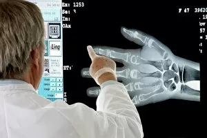
MODEL RELEASED. Radiologist examining a hand X-ray

Background imagePhalanges Collection: Human foot, historical anatomical model

Human foot, historical anatomical model
Human foot, anatomical model. 1965 model showing the skeletal structure of the left human foot and ankle

Background imagePhalanges Collection: Hallux rigidus before surgery, X-ray

Hallux rigidus before surgery, X-ray
Hallux rigidus before surgery. X-ray showing foot bones before surgery to correct a case of hallux rigidus, a stiff big toe

Background imagePhalanges Collection: Forearm muscles, artwork

Forearm muscles, artwork
Forearm muscles. Anatomical computer artwork showing the bones (white), muscles (red), and tendons and ligaments (blue) of a human forearm, wrist and hand

Background imagePhalanges Collection: Ankle anatomy, artwork

Ankle anatomy, artwork
Ankle anatomy. Computer artwork showing the bone (white) of a human foot, and the ligaments (blue) of the ankle. At far right is the Achilles tendon

Background imagePhalanges Collection: Lower leg anatomy, artwork

Lower leg anatomy, artwork
Lower leg anatomy, computer artwork. Front view of a human lower leg and foot, showing the bones (white), muscles (red) and tendons (blue)

Background imagePhalanges Collection: Plantar fascia, artwork

Plantar fascia, artwork
Plantar fascia. Computer artwork of the underside of a human foot, showing the bones and the plantar fascia, a layer of fibrous connective tissue in the sole of the foot that supports the arch

Background imagePhalanges Collection: Human arm musculature, 19th century C015 / 6104

Human arm musculature, 19th century C015 / 6104
Human arm musculature. 19th-century artwork of the anatomy of various arm muscles, including those of the shoulder, arms and hands
Background imagePhalanges Collection: Amputated finger, X-ray C017 / 8010

Amputated finger, X-ray C017 / 8010
Amputated finger. X-ray of the hand of a 17 year old male patient who has had the tip of his middle finger amputated

Background imagePhalanges Collection: Phalanx joint anatomy, artwork C016 / 2884

Phalanx joint anatomy, artwork C016 / 2884
Phalanx joint anatomy. Artwork of a sectioned joint between two phalanx bones (phalanges). These are found in the fingers and toes

Background imagePhalanges Collection: Wrist pain, artwork C013 / 8822

Wrist pain, artwork C013 / 8822
Wrist pain, computer artwork

Background imagePhalanges Collection: Leg ligaments, artwork C013 / 4656

Leg ligaments, artwork C013 / 4656
Leg anatomy. Computer artwork of an anterior view of a human leg, showing the muscles and tendons (brown and white), bones and ligaments (white), arteries and veins (red and blue), nerves (yellow)

Background imagePhalanges Collection: Wrist ligaments, artwork C013 / 4651

Wrist ligaments, artwork C013 / 4651
Wrist ligaments. Computer artwork of the bones of a human hand, showing the ligaments (white) holding together the bones of the wrist

Background imagePhalanges Collection: Thumb muscles, artwork C013 / 4587

Thumb muscles, artwork C013 / 4587
Thumb muscles. Computer artwork of two of the muscles (red) of the thumb and their attachment to the bones of the hand. These are two of the three thenar muscles

Background imagePhalanges Collection: Forearm muscle, artwork C013 / 4585

Forearm muscle, artwork C013 / 4585
Forearm muscle. Computer artwork of an anterior view of a human arm, showing the flexor pollicis longus muscle (red) of the forearm and its attachments to the bones of the arm, wrist and hand


All Professionally Made to Order for Quick Shipping

"Unveiling the Marvels of Phalanges: A Journey into the Intricacies of Human Anatomy" Step into the world of phalanges, where we explore the wonders hidden within our normal foot and its intricate skeletal structure. Peering through an X-ray lens, witness the mesmerizing beauty of a skeleton from below, revealing the delicate balance between bones and joints. Delve deeper with detailed diagrams showcasing the complex network of bones in our hands and arms, unraveling their vital role in everyday activities. Behold a coloured X-ray masterpiece capturing the essence of a healthy man's hand – a true testament to nature's flawless design. Embark on an anatomical adventure as we unveil diagrams depicting the bones that support our right leg and hip, enabling us to stand tall. Explore captivating X-rays that showcase healthy human hands in all their glory – marvel at their intricate composition and functionality. Travel back in time with an Australopithecine or Homo habilis foot cast (OH8), offering insights into our evolutionary journey towards bipedalism. Admire stunning artwork illustrating outer ankle ligaments (C013 / 4452) – discover how these crucial structures provide stability during movement. Immerse yourself further with artwork showcasing inner ankle ligaments (C013 / 4451), shedding light on another aspect of this remarkable joint's anatomy. Witness innocence through a child hand X-ray - see how tiny fingers hold immense potential for growth and exploration. Take a historical leap as we present a vibrant lithograph comparing hand bones across nine different mammals from 1898 - highlighting both similarities and unique adaptations. Thumb your way through life’s intricacies with a colourful lithograph dedicated solely to this essential digit - celebrating its versatility like never before.