Home > Popular Themes > Human Body
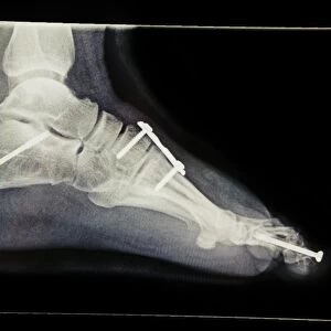
Normal foot, X-ray
![]()

Wall Art and Photo Gifts from Science Photo Library
Normal foot, X-ray
Normal foot. Coloured X-ray of a healthy human foot seen from the top (antero-posterior or dorso- plantar projection). The foot comprises 26 bones and two sesamoid bones (not seen). The toes (top) are formed from phalanx bones. There are two phalanges in the big toe, and three in the other four toes. Behind the toes are five long metatarsal bones (centre). These articulate with the five tarsal bones of the mid-foot. The mid- foot joins the hind-foot at the semi-circular joint (lower centre). The hind-foot comprises the calcaneus (heel) and the talus, which forms the pivot of the ankle with the tibia (bottom)
Science Photo Library features Science and Medical images including photos and illustrations
Media ID 6448075
© DU CANE MEDICAL IMAGING LTD/SCIENCE PHOTO LIBRARY
Ankle Antero Posterior Bones Foot Heel Metatarsal Metatarsals Phalanges Phalanx Radiograph Radiography Tarsal Tarsals Tibia Toes
EDITORS COMMENTS
This print showcases the intricate beauty of a normal human foot, captured through a colored X-ray. From an antero-posterior or dorso-plantar perspective, this image reveals the remarkable complexity and harmony within our feet. Comprising 26 bones and two hidden sesamoid bones, this anatomical marvel is truly awe-inspiring. At first glance, we are drawn to the toes formed by phalanx bones. The big toe stands out with its two phalanges, while the other four toes boast three each. Moving towards the center of the foot, we encounter five elongated metatarsal bones that seamlessly connect with the tarsal bones in the mid-foot region. The mid-foot acts as a crucial junction between these metatarsals and the hind-foot section. Here lies a semi-circular joint that facilitates fluid movement and stability. Speaking of which, let us not forget about our heel – known as calcaneus – which provides essential support during locomotion. Completing this magnificent structure is talus - forming a pivotal point for ankle rotation alongside tibia (shinbone). Together they ensure smooth mobility and weight distribution throughout various activities. This radiograph not only highlights every bone but also serves as an educational tool to understand how our feet function harmoniously on a daily basis. Science Photo Library has once again delivered an extraordinary visual representation of human anatomy - reminding us of both its complexity and elegance.
MADE IN THE UK
Safe Shipping with 30 Day Money Back Guarantee
FREE PERSONALISATION*
We are proud to offer a range of customisation features including Personalised Captions, Color Filters and Picture Zoom Tools
SECURE PAYMENTS
We happily accept a wide range of payment options so you can pay for the things you need in the way that is most convenient for you
* Options may vary by product and licensing agreement. Zoomed Pictures can be adjusted in the Basket.


