
Metatarsals Collection
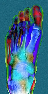
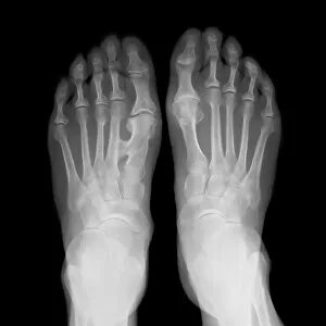
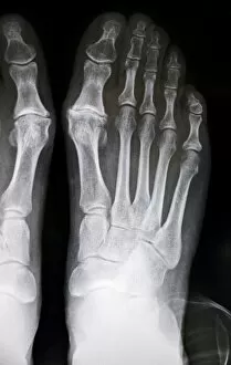
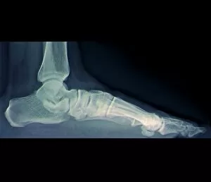
"Exploring the Marvels of Metatarsals: Unveiling the Secrets of Our Feet" A glimpse into a normal foot's intricate structure, as revealed by an X-ray


All Professionally Made to Order for Quick Shipping
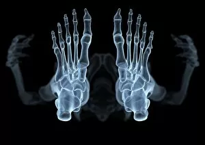
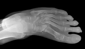
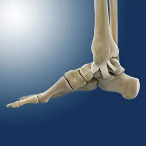
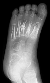
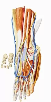
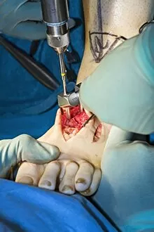
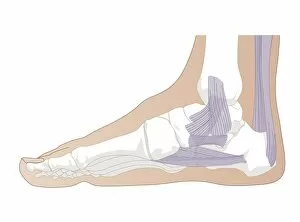
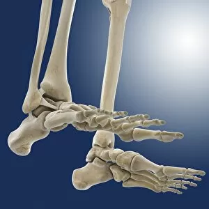
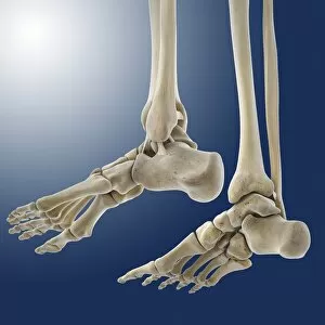
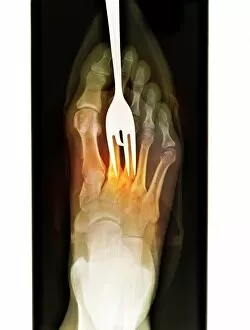
"Exploring the Marvels of Metatarsals: Unveiling the Secrets of Our Feet" A glimpse into a normal foot's intricate structure, as revealed by an X-ray. From below, witness the hidden beauty of our skeletal foundation through an artistic X-ray representation. The haunting image of a Lisfranc fracture captured in an X-ray, reminding us to cherish our metatarsals' resilience. Delicate outer ankle ligaments depicted in stunning artwork (C013 / 4452), showcasing their crucial role in maintaining stability. Dive deeper into the inner ankle ligaments with captivating artwork (C013 / 4451), unraveling their significance for balance and movement. Witness a rare case of fused metatarsals through an intriguing X-ray (C017 / 7168), highlighting the body's remarkable adaptability. An unfortunate broken foot immortalized in an X-ray (C017 / 7975), serving as a reminder to protect and care for our precious metatarsals. Discovering the wonders within: observe a child's foot anatomy through an enlightening X-ray, marveling at its potential for growth and development. Unraveling the complexity: exploring every nook and cranny of foot anatomy to appreciate its incredible functionality and versatility. Embark on a journey through bones that shape us all – uncovering the interconnectedness between metatarsals and other elements within the human skeleton. Peering beneath our skin: unveiling internal foot anatomy to understand how each component contributes to seamless locomotion and support systems. Captivating imagery capturing cavus foot surgery with meticulous metatarsal pinning - celebrating medical advancements that restore mobility. Intriguingly complex yet undeniably fascinating, these glimpses into "metatarsals" offer profound insights into one of nature's most remarkable creations – the human foot.