Home > Popular Themes > Human Body
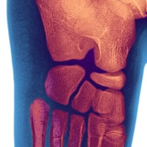
Lisfranc fracture, X-ray

![]()
Wall Art and Photo Gifts from Science Photo Library
Lisfranc fracture, X-ray
Science Photo Library features Science and Medical images including photos and illustrations
Media ID 6325057
© DU CANE MEDICAL IMAGING LTD/SCIENCE PHOTO LIBRARY
Broken Diagnosis Diagnostic Foot Fracture Fractured From Above Injured Injury Joints Metatarsal Metatarsals Radiography Thirties Toes X Ray X Ray Machine Abnormal Displace Mono Chrome Thirty Six Unhealthy
EDITORS COMMENTS
This monochrome print from Science Photo Library showcases the intricate details of a Lisfranc fracture, captured through an X-ray image. The subject of this image is a female in her thirties who unfortunately suffered this painful injury. The photograph highlights the expertise of medical professionals and the power of diagnostic tools like X-ray machines in providing accurate diagnoses. The broken bone, specifically located in the foot's tarsometatarsal articulation or Lisfranc joint, is clearly visible due to its abnormality and displacement. The composition captures the complexity of human anatomy as it zooms in on the injured foot from above. This perspective allows viewers to appreciate both the delicate structure of metatarsals and toes while also emphasizing how severe damage can occur even within these small joints. With its sharp focus and high level of detail, this print serves as a valuable educational resource for medical students, doctors, and researchers studying fractures and injuries related to feet. It offers insight into one specific case – a Lisfranc fracture – but also prompts broader discussions about bone health, diagnosis techniques, and treatment options. Science Photo Library continues to excel at capturing visually striking images that not only inform but also inspire curiosity about our complex bodies and their vulnerabilities.
MADE IN THE UK
Safe Shipping with 30 Day Money Back Guarantee
FREE PERSONALISATION*
We are proud to offer a range of customisation features including Personalised Captions, Color Filters and Picture Zoom Tools
FREE COLORIZATION SERVICE
You can choose advanced AI Colorization for this picture at no extra charge!
SECURE PAYMENTS
We happily accept a wide range of payment options so you can pay for the things you need in the way that is most convenient for you
* Options may vary by product and licensing agreement. Zoomed Pictures can be adjusted in the Basket.