
Diagnosis Collection
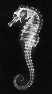
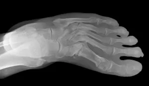
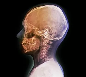
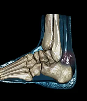
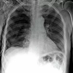
"Unlocking the Secrets: The Power of Diagnosis" A seahorse's hidden world revealed through an X-ray, reminding us that diagnosis goes beyond human health


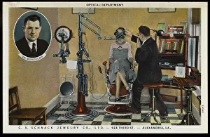


All Professionally Made to Order for Quick Shipping
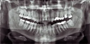
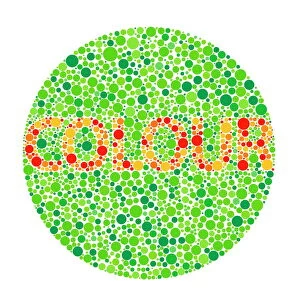
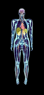
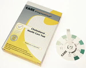
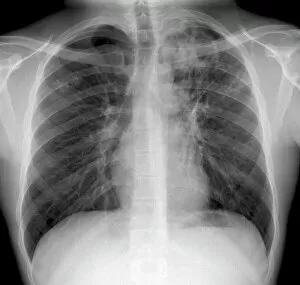
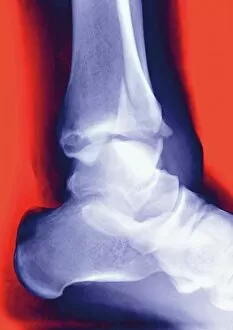
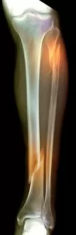
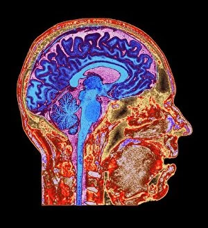
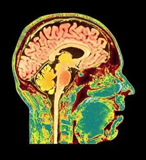
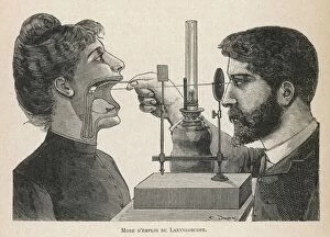
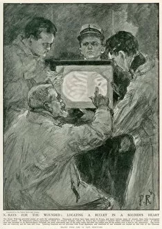
"Unlocking the Secrets: The Power of Diagnosis" A seahorse's hidden world revealed through an X-ray, reminding us that diagnosis goes beyond human health. Unveiling the spectrum: a color blindness test helping us understand how our eyes perceive the world differently. Eyesight test chart guiding us towards clarity and precision in diagnosing visual impairments. Peering into fractures: a broken wrist bone captured on X-ray, aiding doctors in accurate diagnosis and treatment plans. Exploring beneath the surface: a panoramic dental X-ray exposing hidden oral issues for comprehensive diagnoses. Empowering individuals with knowledge: home cholesterol test kits providing early detection for proactive health management. Saving lives through swift intervention: Pneumothorax treatment guided by an X-ray to restore lung function effectively. Knees tell stories too: a normal knee showcased on an X-ray, enabling precise diagnoses of joint conditions and injuries. Delving deeper into mysteries within: full body scans via MRI revealing intricate details for accurate medical assessments. Artistry meets science as ECGs portray the rhythm of a healthy heart, serving as diagnostic masterpieces in healthcare settings. Battling tuberculosis head-on with an X-ray, unveiling its presence so prompt treatments can be initiated promptly