Fraud Blocker Skip to main content

X Ray Machine Collection

The incredible versatility of the x-ray machine never ceases to amaze us

Background imageX Ray Machine Collection: Panoramic dental X-ray

Panoramic dental X-ray showing all the teeth in the upper and lower jaws

Background imageX Ray Machine Collection: Brain blood vessels, 3D angiogram C007 / 1981

Brain blood vessels, 3D angiogram C007 / 1981
Brain blood vessels. Coloured 3D lateral angiogram (blood vessel X-ray) of the blood vessels in the brain of a healthy 32-year-old

Background imageX Ray Machine Collection: Broken wrist bone, X-ray C017 / 7187

Broken wrist bone, X-ray C017 / 7187
Broken wrist bone. X-ray of the hand of a 12 year old male with a fractured trapezium bone. The trapezium is the leftmost bone in the group of bones at the base of the bad

Background imageX Ray Machine Collection: Horses skull

Horses skull, coloured X-ray

Background imageX Ray Machine Collection: Skeleton playing rugby

Skeleton playing rugby. Computer enhanced X- ray of a skeleton running with a rugby ball

Background imageX Ray Machine Collection: Skeleton drinking, X-ray

Skeleton drinking, X-ray
Human skeleton walking. Computer artwork based on an X-ray of a full human skeleton walking. The 206 bones provide protection & structural support, and the joints allow locomotion

Background imageX Ray Machine Collection: Triggerfish skeleton, X-ray

Triggerfish skeleton, X-ray
Triggerfish skeleton. Coloured X-ray of a triggerfish (family Balistidae). Its name originates from its ability to erect the first two dorsal spines (upper left), which can then lock or unlock

Background imageX Ray Machine Collection: Seahorse skeleton

Seahorse skeleton. Coloured x-ray of a seahorse (Hippocampus sp.). Seahorse populations are under threat due to the fishing industry

Background imageX Ray Machine Collection: Horse skull

Horse skull (Equus caballus), side view X-ray

Background imageX Ray Machine Collection: Total hip replacement, X-ray

Total hip replacement, X-ray
Total hip replacement. Coloured frontal X-ray of a female pelvis with a total hip replacement (white, lower right). The replacement hip consists of a ball and shaft

Background imageX Ray Machine Collection: Normal skull, X-ray

Normal skull, X-ray
Normal skull, coloured frontal 3D computed tomography (CT) scan

Background imageX Ray Machine Collection: Primate skulls

Primate skulls. Coloured x-rays of the skulls of a gorilla (Gorilla gorilla, left), chimpanzee (Pan trogoldytes, centre) and human (Homo sapien, left)

Background imageX Ray Machine Collection: Tuberculosis, X-ray

Tuberculosis, X-ray
Tuberculosis. X-ray of the chest of a 25 year old male patient with pulmonary tuberculosis. Affected areas of the lungs (dark areas) are shown by grainy white patches

Background imageX Ray Machine Collection: Person with a camera, X-ray

Person with a camera, X-ray
Person with a camera. Coloured, frontal X-ray of a person holding a camera (blue, left). The persons skull (centre) and neck bones (vertebrae, lower centre) can be seen

Background imageX Ray Machine Collection: Skeleton playing football

Skeleton playing football. Computer enhanced X- ray of a skeleton bouncing a football off his knee

Background imageX Ray Machine Collection: Varicose veins, X-ray

Varicose veins, X-ray
Varicose veins. Coloured X-ray of varicose veins in the calf of a fifty-five-year-old male. The veins (red), which carry blood back to the heart, are swollen, irregular and distorted

Background imageX Ray Machine Collection: Fractured ankle, X-ray

Fractured ankle, X-ray
Fractured ankle. Coloured profile X-ray of a distal fibula fracture (upper centre). The fibula is the smaller leg bone running down from top centre. The larger bone is the tibia

Background imageX Ray Machine Collection: Skeleton playing football

Skeleton playing football. Computer enhanced X- ray of a skeleton kicking a football

Background imageX Ray Machine Collection: Lisfranc fracture, X-ray

Lisfranc fracture, X-ray

Background imageX Ray Machine Collection: Normal spine, X-ray

Normal spine, X-ray
Normal spine. Coloured profile X-ray showing bones of the spine (vertebrae, centre). The front of the body is at right. The ribs (upper right) and the pelvis (bottom) are also seen

Background imageX Ray Machine Collection: Normal neck, X-ray

Normal neck, X-ray
Noraml neck. X-ray of the healthy cervical (neck) spine of a 29 year old male patient

Background imageX Ray Machine Collection: Tension pneumothorax, X-ray

Tension pneumothorax, X-ray
Tension pneumothorax. X-ray of the chest of a patient with tension pneumothorax that has lead to soft-tissue emphysema. Tension pneumothorax is a progressive deterioration

Background imageX Ray Machine Collection: Computer motherboard, coloured X-ray C016 / 7204

Computer motherboard, coloured X-ray C016 / 7204
Computer motherboard, coloured x-ray of the main circuit board (motherboard) of a personal computer (PC). Motherboard components include transistors, diodes, resistors, capacitors and inductors

Background imageX Ray Machine Collection: Fractured jawbone, X-ray

Fractured jawbone, X-ray
Fractured jawbone. Coloured X-ray of a human jaw showing a fractured lower jawbone (mandible). The fracture has occurred to the left side of the lower jawbone (bottom right, orange)

Background imageX Ray Machine Collection: Mobile phone use, X-ray

Mobile phone use, X-ray. The person at right is showing the other a picture (of a skull) on the mobile phone

Background imageX Ray Machine Collection: X-ray security machine, 1900

X-ray security machine, 1900
X-ray security machine. Historical artwork of a fluoroscope being used in a late 19th-century French customs hall. The fluoroscope was designed and built by the French inventor Eugene Ducretet

Background imageX Ray Machine Collection: Turtle

Turtle, coloured X-ray. The head of the turtle is at right

Background imageX Ray Machine Collection: Neck, side view, coloured X-ray

Neck, side view, coloured X-ray
Neck from the side, coloured X-ray. The front of the body is at right. The spine runs down centre. It is made up of block-like bones called vertebrae

Background imageX Ray Machine Collection: Spring train, X-ray

Spring train, X-ray
Spring train. X-ray showing the mechanism inside a toy train

Background imageX Ray Machine Collection: Spring train, X-ray

Spring train, X-ray
Spring train. X-ray showing the mechanism inside a toy train

Background imageX Ray Machine Collection: Broken ankle, X-ray C017 / 7185

Broken ankle, X-ray C017 / 7185
Broken ankle. X-ray of the lower leg of an 85 year old female with fractures at the ends of the tibia (shin bone, right) and fibula (calf bone, left)

Background imageX Ray Machine Collection: Amputated lower leg, X-ray C017 / 7644

Amputated lower leg, X-ray C017 / 7644
Amputated lower leg, X-ray. The leg has been amputated below the knee. The tops of the tibia (shin bone, lower centre) and fibula (calf bone, lower right) are seen

Background imageX Ray Machine Collection: Asbestosis, X-ray C017 / 7143

Asbestosis, X-ray C017 / 7143
Asbestosis. X-ray of the chest of a 78 year old male that has been exposed to asbestos, showing diffuse plaques (light grey areas) in the lungs (dark)

Background imageX Ray Machine Collection: Fused metatarsals, X-ray C017 / 7168

Fused metatarsals, X-ray C017 / 7168
Fused metatarsals. X-ray of the feet of a 73 year old female patient showing the fusing the the big toes and second toe metatarsal bones (mid foot bones) in the left foot

Background imageX Ray Machine Collection: Pneumothorax, X-ray C017 / 7148

Pneumothorax, X-ray C017 / 7148
Pneumothorax. X-ray of the chest of a 50 year old male patient with a pneumothorax, or collapsed lung. The right lung (left on X-ray) has collapsed due to a build-up of air (solid grey)

Background imageX Ray Machine Collection: Arthritis of the neck, X-ray C017 / 7389

Arthritis of the neck, X-ray C017 / 7389
Arthritis of the neck. X-ray of the neck of a 67 year old female with osteoarthritis of the neck. Osteoarthritis of the neck, also known as cervical spondylosis

Background imageX Ray Machine Collection: Normal legs, X-rays

Normal legs, X-rays
Normal legs. Coloured frontal X-rays of the healthy legs, of a 35-year-old

Background imageX Ray Machine Collection: Computer motherboard, coloured X-ray C016 / 7212

Computer motherboard, coloured X-ray C016 / 7212
Computer motherboard, coloured x-ray of the main circuit board (motherboard) of a personal computer (PC). Motherboard components include transistors, diodes, resistors, capacitors and inductors

Background imageX Ray Machine Collection: Broken leg, X-ray C017 / 7977

Broken leg, X-ray C017 / 7977
Broken leg. X-ray of the leg of a 32 year old male who has been in a parachuting accident. The fibula (calf bone, right) is fractured

Background imageX Ray Machine Collection: Broken foot, X-ray C017 / 7975

Broken foot, X-ray C017 / 7975
Broken foot. X-ray of the foot of a 3 year old male patient with fractured second and third metatarsals (foot bones)

Background imageX Ray Machine Collection: Pinned broken arm, X-ray C017 / 7869

Pinned broken arm, X-ray C017 / 7869
Pinned broken arm. X-ray of the arm of a patient that has been internally fixed after fracturing both of the lower arm bones, the ulna and radius

Background imageX Ray Machine Collection: Broken arm, X-ray C017 / 7863

Broken arm, X-ray C017 / 7863
Broken arm. X-ray of the arm of a patient that has broken both of the lower arm bones, the ulna and radius

Background imageX Ray Machine Collection: Ostrich skeleton

Ostrich skeleton. Coloured x-ray of an ostrich (Struthio camelus). This bird is native to Africa and it is the largest species of bird

Background imageX Ray Machine Collection: Adult human skull

Adult human skull. Side view X-ray showing the cranium, eye socket, nasal area and teeth. For an X-ray of a babys skull see image P120/223 and for an X-ray of a childs skull see image number P120/224

Background imageX Ray Machine Collection: Broken arm bone, digital X-ray

Broken arm bone, digital X-ray
Broken arm bone. Digital frontal X-ray of the fractured right humerus (upper arm bone) of a patient in their early twenties

Background imageX Ray Machine Collection: Normal hand, digital X-ray

Normal hand, digital X-ray
Normal hand. Digital X-ray of the normal right hand of a patient in their late twenties. Various sets of data relating to the image can be seen around the edges of the X-ray

Background imageX Ray Machine Collection: Broken arm, X-ray

Broken arm, X-ray
Broken arm. X-ray of the forearm of a 29 year old male patient with a fractured ulna. Treatment is with immobilisation of the bone, either externally using a cast, or internally using pins

Background imageX Ray Machine Collection: Ischaemia, digital angiogram

Ischaemia, digital angiogram
image d angiographie numerisee( non soustraite) de la main gauche, en vue de face, par catheterisme hyperselectif des arteres du membre superieur gauche, sous anasthesie generale


All Professionally Made to Order for Quick Shipping

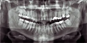
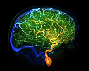
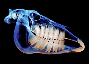
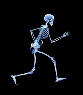
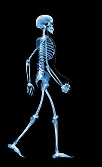
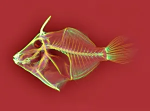
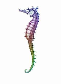
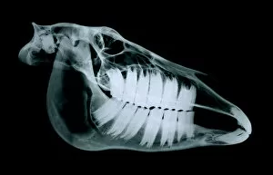
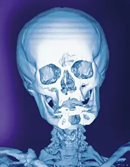
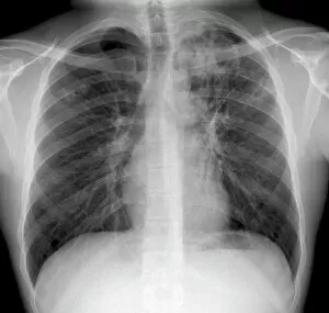
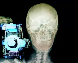
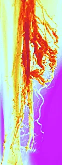
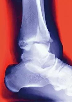
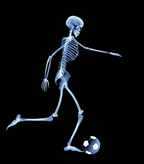
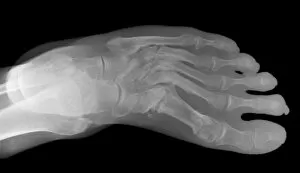
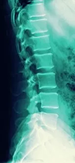
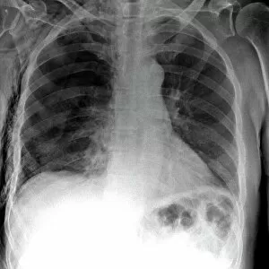
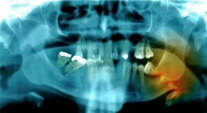
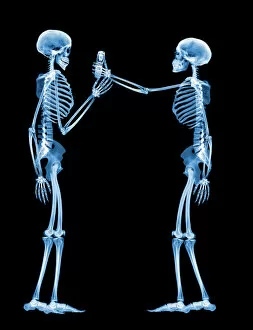
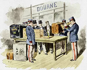
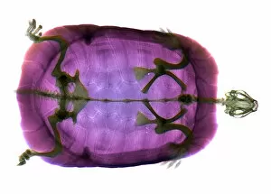
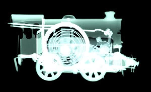
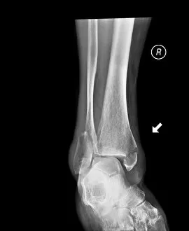
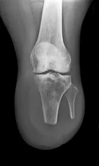
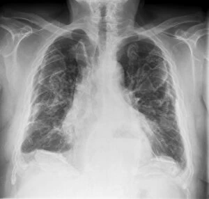
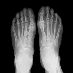
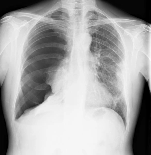
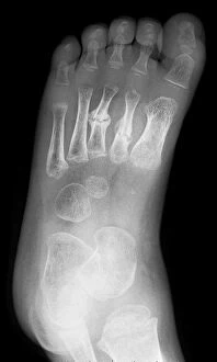
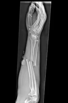
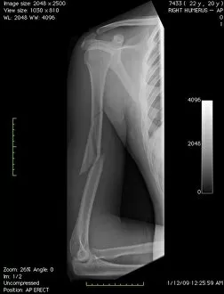
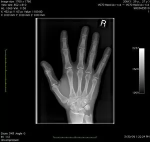
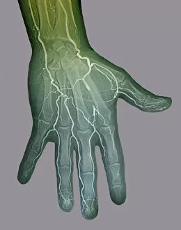
The incredible versatility of the x-ray machine never ceases to amaze us. From capturing intricate details of brain blood vessels in a 3D angiogram C007 / 1981, to diagnosing a broken wrist bone in X-ray C017 / 7187, this technology has revolutionized medical imaging. But it doesn't stop there. The x-ray machine has even ventured into the animal kingdom, revealing fascinating insights such as a horse's skull or the delicate skeleton of a seahorse. It seems that no creature is too small or too large for this remarkable tool. In addition to its medical applications, the x-ray machine also showcases its playful side. Who would have thought that skeletons could engage in sports? A rugby-playing skeleton and even one enjoying a refreshing drink are just some how this technology can bring humor and creativity to our lives. Not limited to humans and animals, the x-ray machine extends its reach into other fields as well. Panoramic dental X-rays help dentists assess oral health with precision while an individual holding a camera reveals how this invention itself can be captured on film. Furthermore, we witness how the x-ray machine aids in surgical procedures like total hip replacements by providing surgeons with detailed images for precise placement and alignment. Last but not least, let's not forget about exploring what lies beneath our own skin - normal skulls are examined through X-rays allowing us to marvel at the intricacies hidden within each one of us. From healthcare advancements to artistic expressions and scientific discoveries, these glimpses into various aspects of life demonstrate why the x-ray machine remains an invaluable tool that continues to shape our understanding of both human anatomy and beyond.