Fraud Blocker Skip to main content

Home > Science > Xray

Xray Collection

X-rays are a type of electromagnetic radiation that have shorter wavelengths than visible light

Choose a picture from our Xray Collection for your Wall Art and Photo Gifts

547 items

Background imageXray Collection: Xray X-MP / 48 supercomputer at CERN

Xray X-MP / 48 supercomputer at CERN
The distinctive shape & bright colouring of the CRAY X-MP/48 supercomputer dominate the Computer Centre at CERN, the European centre for particle physics research near Geneva

Background imageXray Collection: Brain blood vessels, 3D angiogram C007 / 1981

Brain blood vessels, 3D angiogram C007 / 1981
Brain blood vessels. Coloured 3D lateral angiogram (blood vessel X-ray) of the blood vessels in the brain of a healthy 32-year-old

Background imageXray Collection: Amputated lower leg, X-ray C017 / 7644

Amputated lower leg, X-ray C017 / 7644
Amputated lower leg, X-ray. The leg has been amputated below the knee. The tops of the tibia (shin bone, lower centre) and fibula (calf bone, lower right) are seen

Background imageXray Collection: Broken ankle, X-ray C017 / 7185

Broken ankle, X-ray C017 / 7185
Broken ankle. X-ray of the lower leg of an 85 year old female with fractures at the ends of the tibia (shin bone, right) and fibula (calf bone, left)

Background imageXray Collection: Broken wrist bone, X-ray C017 / 7187

Broken wrist bone, X-ray C017 / 7187
Broken wrist bone. X-ray of the hand of a 12 year old male with a fractured trapezium bone. The trapezium is the leftmost bone in the group of bones at the base of the bad

Background imageXray Collection: Normal childs head, X-ray

Normal childs head, X-ray
Normal childs head. Coloured X-ray of the head of a 13 year old

Background imageXray Collection: X-ray Stiletto High-Heeled Shoe

X-ray Stiletto High-Heeled Shoe
Coloured X-ray of a female leg wearing Stiletto High-Heeled Shoe

Background imageXray Collection: Computer motherboard, coloured X-ray C016 / 7204

Computer motherboard, coloured X-ray C016 / 7204
Computer motherboard, coloured x-ray of the main circuit board (motherboard) of a personal computer (PC). Motherboard components include transistors, diodes, resistors, capacitors and inductors

Background imageXray Collection: Asbestosis, X-ray C017 / 7143

Asbestosis, X-ray C017 / 7143
Asbestosis. X-ray of the chest of a 78 year old male that has been exposed to asbestos, showing diffuse plaques (light grey areas) in the lungs (dark)

Background imageXray Collection: Fused metatarsals, X-ray C017 / 7168

Fused metatarsals, X-ray C017 / 7168
Fused metatarsals. X-ray of the feet of a 73 year old female patient showing the fusing the the big toes and second toe metatarsal bones (mid foot bones) in the left foot

Background imageXray Collection: Pneumothorax, X-ray C017 / 7148

Pneumothorax, X-ray C017 / 7148
Pneumothorax. X-ray of the chest of a 50 year old male patient with a pneumothorax, or collapsed lung. The right lung (left on X-ray) has collapsed due to a build-up of air (solid grey)

Background imageXray Collection: Arthritis of the neck, X-ray C017 / 7389

Arthritis of the neck, X-ray C017 / 7389
Arthritis of the neck. X-ray of the neck of a 67 year old female with osteoarthritis of the neck. Osteoarthritis of the neck, also known as cervical spondylosis

Background imageXray Collection: Normal legs, X-rays

Normal legs, X-rays
Normal legs. Coloured frontal X-rays of the healthy legs, of a 35-year-old

Background imageXray Collection: Salivary gland stone, X-ray

Salivary gland stone, X-ray
Salivary gland stone. X-ray of the jaw of a 36-year-old patient with a stone (white, lower left) in the right submaxillary gland

Background imageXray Collection: Computer motherboard, coloured X-ray C016 / 7212

Computer motherboard, coloured X-ray C016 / 7212
Computer motherboard, coloured x-ray of the main circuit board (motherboard) of a personal computer (PC). Motherboard components include transistors, diodes, resistors, capacitors and inductors

Background imageXray Collection: Broken leg, X-ray C017 / 7977

Broken leg, X-ray C017 / 7977
Broken leg. X-ray of the leg of a 32 year old male who has been in a parachuting accident. The fibula (calf bone, right) is fractured

Background imageXray Collection: Broken foot, X-ray C017 / 7975

Broken foot, X-ray C017 / 7975
Broken foot. X-ray of the foot of a 3 year old male patient with fractured second and third metatarsals (foot bones)

Background imageXray Collection: Pinned broken arm, X-ray C017 / 7869

Pinned broken arm, X-ray C017 / 7869
Pinned broken arm. X-ray of the arm of a patient that has been internally fixed after fracturing both of the lower arm bones, the ulna and radius

Background imageXray Collection: Broken arm, X-ray C017 / 7863

Broken arm, X-ray C017 / 7863
Broken arm. X-ray of the arm of a patient that has broken both of the lower arm bones, the ulna and radius

Background imageXray Collection: Ischaemia, digital angiogram

Ischaemia, digital angiogram
image d angiographie numerisee( non soustraite) de la main gauche, en vue de face, par catheterisme hyperselectif des arteres du membre superieur gauche, sous anasthesie generale

Background imageXray Collection: Fractured vertebra, X-ray C017 / 7586

Fractured vertebra, X-ray C017 / 7586
Fractured vertebra. X-ray of the lower spine of a 50 year old male with a fracture to the L1 lumbar vertebra (thord block from top)

Background imageXray Collection: Deformed hands, X-ray C017 / 7561

Deformed hands, X-ray C017 / 7561
Deformed hands. X-ray of the hands of a 7 year old female patient with congenital (present at birth) abnormalities of the fingers

Background imageXray Collection: Hip fracture surgery C017 / 1973

Hip fracture surgery C017 / 1973
MODEL RELEASED. Hip fracture surgery. Radiographer checking an X0ray of a patient with a fractured hip during surgery to fix the hip. A needle can be seen inserted into the hip on the X-ray

Background imageXray Collection: Fish hook in finger, X-ray C017 / 7160

Fish hook in finger, X-ray C017 / 7160
Fish hook. X-ray of a fish hook stuck in a 22 year old males finger

Background imageXray Collection: Pinned boken ankle, X-ray C017 / 7186

Pinned boken ankle, X-ray C017 / 7186
Pinned broken ankle. X-ray of the lower leg of an 85 year old female with fractures at the ends of the tibia (shin bone, right) and fibula (calf bone, left)

Background imageXray Collection: Swallowed key and blades, X-ray C017 / 7559

Swallowed key and blades, X-ray C017 / 7559
Swallowed key and blades. X-ray of the abdomen of a 81 year old male patient that has swallowed several blades and a key (lower left)

Background imageXray Collection: Amputated toes, X-ray C017 / 7166

Amputated toes, X-ray C017 / 7166
Amputated toe. X-ray of the left foot of a 75 year old male patient where the tip of the big toe and two thirds of the next toe have been amputated

Background imageXray Collection: Broken jaw, X-ray C017 / 7557

Broken jaw, X-ray C017 / 7557
Broken jaw. X-ray of the jaw of a 22 year old male patient with a fractured mandible (lower jaw bone). The fracture is seen at left

Background imageXray Collection: Digital SLR camera, coloured X-ray F007 / 4204

Digital SLR camera, coloured X-ray F007 / 4204
Digital SLR (Singe Lens Reflex) camera, coloured x-ray

Background imageXray Collection: Arthrosis of the hand, X-ray F006 / 4616

Arthrosis of the hand, X-ray F006 / 4616
Coloured X-ray of the hand of a patient with arthrosis, particularly the saddle joint. Arthrosis is a degenerative joint disease that damages cartilage and bone

Background imageXray Collection: Foreign object in rectum, X-ray F008 / 3480

Foreign object in rectum, X-ray F008 / 3480
Foreign object in rectum. Coloured X-ray of a foreign object in the rectum of a 31 year old male

Background imageXray Collection: Lung, X-ray F008 / 3218

Lung, X-ray F008 / 3218
Lung. Coloured X-ray showing the blood vessels in a lung

Background imageXray Collection: Amputated fingers, X-ray C017 / 7150

Amputated fingers, X-ray C017 / 7150
Amputated fingers. X-ray of the right hand of a 57 year old male patient where the tips of the fourth and fifth fingers have been amputated

Background imageXray Collection: Radiologist examining knee MRI scans C014 / 1300

Radiologist examining knee MRI scans C014 / 1300
MODEL RELEASED. Radiologist examining knee MRI (magnetic resonance imaging) scans

Background imageXray Collection: Healthy knee, X-ray F006 / 9125

Healthy knee, X-ray F006 / 9125
Healthy knee. Coloured frontal (right) and profile (left) X-rays of the healthy knee of a 24 year old patient

Background imageXray Collection: X-ray of a couple kissing

X-ray of a couple kissing

Background imageXray Collection: Brain aneurysm, 3D scan

Brain aneurysm, 3D scan
Brain aneurysm. 3D computed tomography (CT) angiogram coupled with a magnetic resonance imaging (MRI) scan of the brain of a 38-year-old, showing a large aneurysm (bright)

Background imageXray Collection: Thinking man with a lightbulb

Thinking man with a lightbulb
X-ray of a thinking man with a lightbulb

Background imageXray Collection: Scoliosis of the spine, X-ray C016 / 6566

Scoliosis of the spine, X-ray C016 / 6566
Scoliosis of the spine. Coloured X-ray of the chest of a 34 year old female patient with scoliosis (sideways curvature) of the spine

Background imageXray Collection: Throat cancer, X-rays

Throat cancer, X-rays
Throat cancer. Coloured X-rays showing the transit of a contrast medium (dense white) through the throat of a 54-year-old patient with an adenocarcinoma (cancer) of the lower oesophagus (gullet)

Background imageXray Collection: Human head, X-ray artwork

Human head, X-ray artwork
Human head. Computer artwork of an X-ray through a human head, showing the brain (grey, top), cerebellum (blue), and brainstem and spinal cord (green)

Background imageXray Collection: Balloon angioplasty, X-ray

Balloon angioplasty, X-ray
Balloon angioplasty. Coloured X-ray of the abdomen of a 45-year-old patient with a history of thrombosis (blood clots) in the inferior vena cava (vein), which have narrowed (stenosed)

Background imageXray Collection: External carotid artery, 3D CT scan C016 / 6341

External carotid artery, 3D CT scan C016 / 6341
External carotid artery. Coloured 3D computed tomography (CT) angiogram (blood vessel scan) of the head showing a normal external carotid artery (red, in neck) and its branches

Background imageXray Collection: Healthy knee, CT scan C018 / 0413

Healthy knee, CT scan C018 / 0413
Healthy knee. Coloured frontal computed tomography (CT) scan projected on to a magnetic resonance imaging (MRI) scan of the knee of a 30 year old

Background imageXray Collection: Heart and lung disease, X-ray C018 / 0498

Heart and lung disease, X-ray C018 / 0498
Heart and lung disease. Coloured X-ray of the chest of an 80 year old patient with heart and lung disease. The patient is a former miner and has chronic obstructive pulmonary disease (COPD)

Background imageXray Collection: Broken ribs, X-ray C017 / 7566

Broken ribs, X-ray C017 / 7566
Broken ribs. X-ray of the chest of a 85 year old male patient with fracture to his right ribs (seen on left)

Background imageXray Collection: Artery damage, MRA scan

Artery damage, MRA scan
Artery damage. Magnetic resonance angiography (MRA) scan of the neck of a 23-year-old male motorcycle crash victim. Dissection of the right internal carotid artery (left)

Background imageXray Collection: Computer motherboard, X-ray C016 / 7208

Computer motherboard, X-ray C016 / 7208
Computer motherboard, x-ray of the main circuit board (motherboard) of a personal computer (PC). Motherboard components include transistors, diodes, resistors, capacitors and inductors


All Professionally Made to Order for Quick Shipping


-

Xray Collection

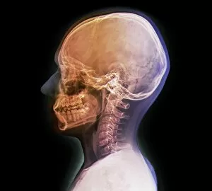
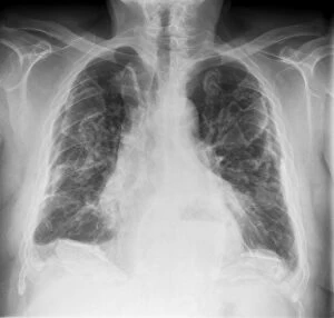
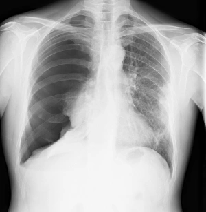
X-rays are a type of electromagnetic radiation that have shorter wavelengths than visible light. They were discovered in 1895 by German physicist Wilhelm Conrad Röntgen, who found that they could penetrate through solid objects and produce images on photographic plates. X-rays are used extensively in medical imaging to diagnose and treat various conditions, such as broken bones, tumors, and dental problems and can also be used for non-medical purposes, such as studying the structure of materials or detecting hidden defects in manufactured products. However, exposure to high levels of X-rays can be harmful to human health due to their ionizing nature.
+

Our beautiful pictures are available as Framed Prints, Photos, Wall Art and Photo Gifts

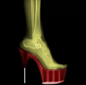
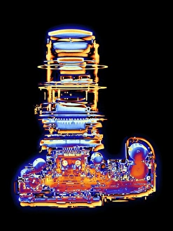
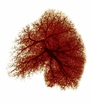
The Xray collection from Media Storehouse offers a unique and fascinating glimpse into the world of science. Our collection features stunning images of various objects, animals, and even humans captured using x-ray technology. The intricate details and inner workings of these subjects are revealed in a way that is both beautiful and informative. From the delicate bones of birds to the complex machinery inside industrial equipment, our collection showcases the incredible versatility of x-ray imaging. These prints make for striking wall art or framed pieces that will add an element of curiosity to any room. Whether you're a science enthusiast or simply appreciate visually stunning artwork, the Xray collection from Media Storehouse is sure to impress. With high-quality prints available in a range of sizes, there's something for everyone in this captivating collection.
+

What are Xray (Science) art prints?

Xray art prints are a unique and fascinating collection of images that showcase the beauty and complexity of the natural world. These prints feature X-ray photographs of various objects, from flowers to animals, revealing their intricate inner workings in stunning detail. The use of X-rays allows us to see beyond what is visible to the naked eye, providing a new perspective on familiar subjects. These art prints are perfect for those who appreciate science and nature, as well as anyone looking for something truly unique to decorate their home or office. They make great conversation starters and can be used to add interest and depth to any space. We offer a wide range of Xray art prints in different sizes and formats, including framed options for easy display. Each print is produced using high-quality materials to ensure long-lasting durability and vibrant colors that will stand out wherever they are displayed.
+

What Xray (Science) art prints can I buy from Media Storehouse?

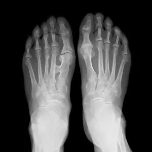
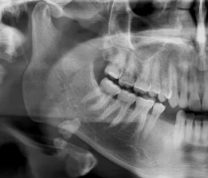
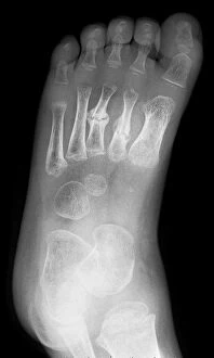
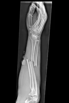
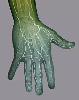
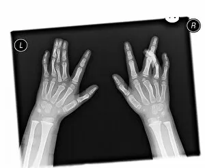
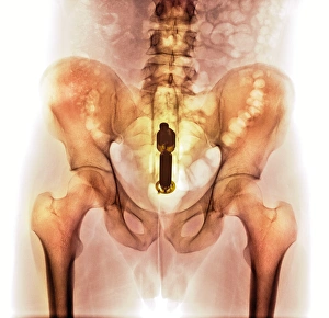
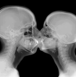
At Media Storehouse, you can find a wide range of Xray art prints that are perfect for adding a unique touch to your home or office decor. These prints showcase the intricate details and inner workings of various objects, from flowers and animals to machines and tools. Some popular options include X-ray images of human bones, such as hands and feet, which make for interesting conversation starters in medical offices or clinics. You can also find stunning X-ray photographs of flowers like tulips or roses that reveal their delicate structures in an entirely new way. For those interested in technology, there are plenty of X-ray images available showcasing the inner workings of gadgets like cameras or watches. And if you're looking for something truly unique, consider an X-ray print featuring a vintage car engine or even a musical instrument like a guitar. No matter what your interests may be, we have plenty of beautiful and fascinating Xray art prints to choose from.
+

How do I buy Xray (Science) art prints?

To buy Xray art prints from Media Storehouse, you can browse through our extensive collection of images featuring various scientific subjects. Once you have found the print that you like, simply select the size and format that suits your needs and add it to your cart. We offer a range of formats for their prints, including canvas, framed prints, and posters. You can also choose from different sizes depending on where you plan to display the artwork. Once you have added all the items to your cart, proceed to checkout and follow the prompts to complete your purchase. Payment options may vary depending on your location and preferred method of payment. Buying Xray art prints from Media Storehouse is a straightforward process that allows you to easily find and purchase high-quality artwork for personal or commercial use.
+

How much do Xray (Science) art prints cost?

We offer a wide range of Xray art prints that vary in price depending on the size and type of print you choose. Our collection includes high-quality photographic prints, canvas prints, framed prints, and more. We understand that everyone has different preferences when it comes to artwork, which is why we strive to provide a diverse selection at affordable prices. Our Xray art prints are perfect for adding a unique touch to any room in your home or office. Whether you're looking for something bold and colorful or subtle and minimalist, our collection has something for everyone. We take pride in offering high-quality products that are both beautiful and durable. If you're interested in purchasing one of our Xray art prints but aren't sure where to start, don't hesitate to reach out to us. Our team is always happy to help customers find the perfect piece of artwork for their space.
+

How will my Xray (Science) art prints be delivered to me?

We take great care in ensuring that your Xray art prints are delivered to you safely and securely. We use high-quality packaging materials to protect your artwork during transit and ensure it arrives in pristine condition. Your prints will be carefully rolled and placed into a sturdy cardboard tube for delivery. This method of shipping ensures that your artwork is protected from any damage or creases during transportation. We work with trusted courier partners who offer reliable delivery services worldwide. Once your order has been dispatched, you will receive an email notification with tracking information so you can follow the progress of your package. We understand how important it is to receive your artwork promptly and in perfect condition. That's why we take every step necessary to ensure that our customers receive their orders quickly and securely.