Premium Framed Print > Science > SEM
Premium Framed Print : Uterus lining during menstruation, SEM


![]()
Framed Photos from Science Photo Library
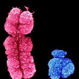
Uterus lining during menstruation, SEM
Uterus during menstruation. Coloured scanning electron micrograph (SEM) of the lining of the uterus being shed during menstruation. The upper layers (red) are being shed and the breakdown of the underlying blood vessels releases red blood cells (red dots). Menstruation occurs for a few days during a womans menstrual cycle. Prior to menstruation, the uterine lining (endometrium) thickens to prepare it for the reception of a fertilised egg. If a fertilised egg enters the uterus, it implants in the wall and develops into an embryo. If the released egg is not fertilised, the thickened wall is shed
Science Photo Library features Science and Medical images including photos and illustrations
Media ID 6449809
© STEVE GSCHMEISSNER/SCIENCE PHOTO LIBRARY
Blood Epithelium Erythrocyte Erythrocytes Lining Magnified Image Menstruation Microscopic Subjects Period Physiological Physiology Re Production Red Blood Cells Reproductive System Shedding Tissue Uterine Uterus Womb False Coloured
17"x15" (43x38cm) Premium Frame
FSC real wood frame with double mounted 10x8 print. Double mounted with white conservation mountboard. Frame moulding comprises stained composite natural wood veneers (Finger Jointed Pine) 39mm wide by 21mm thick. Archival quality Fujifilm CA photo paper mounted onto 1mm card. Overall outside dimensions are 17x15 inches (431x381mm). Rear features Framing tape to cover staples, 50mm Hanger plate, cork bumpers. Glazed with durable thick 2mm Acrylic to provide a virtually unbreakable glass-like finish. Acrylic Glass is far safer, more flexible and much lighter than typical mineral glass. Moreover, its higher translucency makes it a perfect carrier for photo prints. Acrylic allows a little more light to penetrate the surface than conventional glass and absorbs UV rays so that the image and the picture quality doesn't suffer under direct sunlight even after many years. Easily cleaned with a damp cloth. Please note that, to prevent the paper falling through the mount window and to prevent cropping of the original artwork, the visible print may be slightly smaller to allow the paper to be securely attached to the mount without any white edging showing and to match the aspect ratio of the original artwork.
FSC Real Wood Frame and Double Mounted with White Conservation Mountboard - Professionally Made and Ready to Hang
Estimated Image Size (if not cropped) is 24.4cm x 18.3cm (9.6" x 7.2")
Estimated Product Size is 43.1cm x 38.1cm (17" x 15")
These are individually made so all sizes are approximate
Artwork printed orientated as per the preview above, with landscape (horizontal) orientation to match the source image.
FEATURES IN THESE COLLECTIONS
EDITORS COMMENTS
This print from Science Photo Library provides a close-up view of the uterus lining during menstruation. The image, captured using a scanning electron microscope (SEM), reveals the intricate details of this physiological process that occurs in women's bodies. In the image, we can observe the upper layers of the uterine lining being shed, depicted in vibrant red hues. As these layers break down, underlying blood vessels release numerous red blood cells, represented by tiny red dots scattered throughout the image. This shedding and release of blood cells signify menstruation – a natural occurrence lasting for several days during a woman's menstrual cycle. Prior to menstruation, the uterine lining thickens in preparation for potential fertilization and implantation of an egg. If fertilization does occur, an embryo develops within the uterus wall. However, if no fertilized egg is present, this thickened wall is shed as part of menstruation. The SEM technique used to capture this magnified image allows us to appreciate both its biological beauty and scientific significance. It offers valuable insights into reproductive anatomy and physiology while highlighting key elements such as erythrocytes (red blood cells) and epithelium. This remarkable photograph not only showcases the complexity of female reproductive biology but also serves as a reminder of how our bodies undergo fascinating processes to support human life.
MADE IN THE UK
Safe Shipping with 30 Day Money Back Guarantee
FREE PERSONALISATION*
We are proud to offer a range of customisation features including Personalised Captions, Color Filters and Picture Zoom Tools
SECURE PAYMENTS
We happily accept a wide range of payment options so you can pay for the things you need in the way that is most convenient for you
* Options may vary by product and licensing agreement. Zoomed Pictures can be adjusted in the Basket.