Framed Print > Science > SEM
Framed Print : Uterus lining during menstruation, SEM


![]()
Framed Photos from Science Photo Library
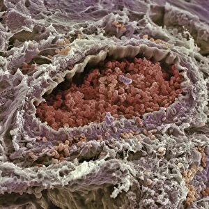
Uterus lining during menstruation, SEM
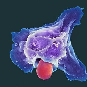
Uterus during menstruation. Coloured scanning electron micrograph (SEM) of the lining of the uterus being shed during menstruation. The upper layers (red) are being shed and the breakdown of the underlying blood vessels releases red blood cells (red dots). Menstruation occurs for a few days during a womans menstrual cycle. Prior to menstruation, the uterine lining (endometrium) thickens to prepare it for the reception of a fertilised egg. If a fertilised egg enters the uterus, it implants in the wall and develops into an embryo. If the released egg is not fertilised, the thickened wall is shed
Science Photo Library features Science and Medical images including photos and illustrations
Media ID 6449809
© STEVE GSCHMEISSNER/SCIENCE PHOTO LIBRARY
Blood Epithelium Erythrocyte Erythrocytes Lining Magnified Image Menstruation Microscopic Subjects Period Physiological Physiology Re Production Red Blood Cells Reproductive System Shedding Tissue Uterine Uterus Womb False Coloured
14"x12" (38x32cm) Modern Frame
Discover the intricacies of the human body with our captivating Framed Prints from Media Storehouse. This particular piece showcases a mesmerizing Coloured Scanning Electron Micrograph (SEM) image of the uterus lining during menstruation, courtesy of Science Photo Library. Delve deeper into the wonders of biology and enhance your space with this educational and visually striking addition. Experience the beauty of science in your home or office.
Wood effect frame, card mounted, 10x8 archival quality photo print. Overall outside dimensions 14x12 inches (38x32cm). Environmentally and ozone friendly, 40mm wide x 15mm Polycore® moulding has the look of real wood, is durable and light and easy to hang. Biodegradable and made with non-chlorinated gases (no toxic fumes) it is efficient; producing 100 tons of polystyrene can save 300 tons of trees! Prints are glazed with lightweight, shatterproof, optical clarity acrylic (providing the same general protection from the environment as glass). The back is stapled hardboard with a sawtooth hanger attached. Note: To minimise original artwork cropping, for optimum layout, and to ensure print is secure, the visible print may be marginally smaller
Contemporary Framed and Mounted Prints - Professionally Made and Ready to Hang
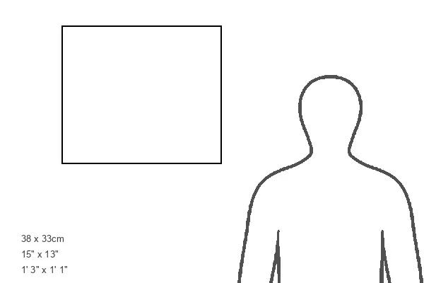
Estimated Image Size (if not cropped) is 24.4cm x 18.3cm (9.6" x 7.2")
Estimated Product Size is 37.6cm x 32.5cm (14.8" x 12.8")
These are individually made so all sizes are approximate
Artwork printed orientated as per the preview above, with landscape (horizontal) orientation to match the source image.
FEATURES IN THESE COLLECTIONS
EDITORS COMMENTS
This print from Science Photo Library provides a close-up view of the uterus lining during menstruation. The image, captured using a scanning electron microscope (SEM), reveals the intricate details of this physiological process that occurs in women's bodies. In the image, we can observe the upper layers of the uterine lining being shed, depicted in vibrant red hues. As these layers break down, underlying blood vessels release numerous red blood cells, represented by tiny red dots scattered throughout the image. This shedding and release of blood cells signify menstruation – a natural occurrence lasting for several days during a woman's menstrual cycle. Prior to menstruation, the uterine lining thickens in preparation for potential fertilization and implantation of an egg. If fertilization does occur, an embryo develops within the uterus wall. However, if no fertilized egg is present, this thickened wall is shed as part of menstruation. The SEM technique used to capture this magnified image allows us to appreciate both its biological beauty and scientific significance. It offers valuable insights into reproductive anatomy and physiology while highlighting key elements such as erythrocytes (red blood cells) and epithelium. This remarkable photograph not only showcases the complexity of female reproductive biology but also serves as a reminder of how our bodies undergo fascinating processes to support human life.
MADE IN THE UK
Safe Shipping with 30 Day Money Back Guarantee
FREE PERSONALISATION*
We are proud to offer a range of customisation features including Personalised Captions, Color Filters and Picture Zoom Tools
SECURE PAYMENTS
We happily accept a wide range of payment options so you can pay for the things you need in the way that is most convenient for you
* Options may vary by product and licensing agreement. Zoomed Pictures can be adjusted in the Basket.