
Microscopic Subjects Collection
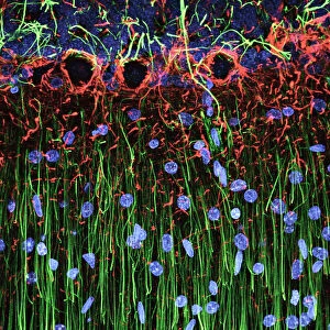
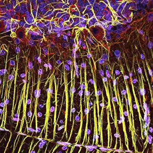
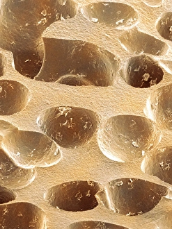
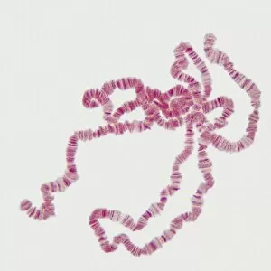
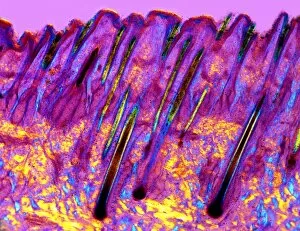
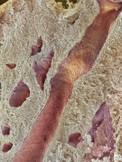
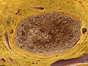
"Unveiling the Unseen: Exploring Microscopic Marvels" Delving into the intricate world of cerebellum tissue


All Professionally Made to Order for Quick Shipping
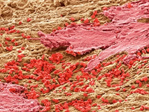
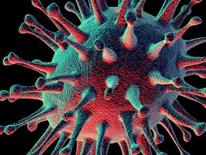
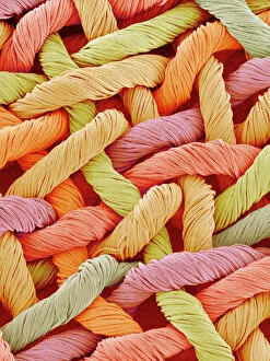
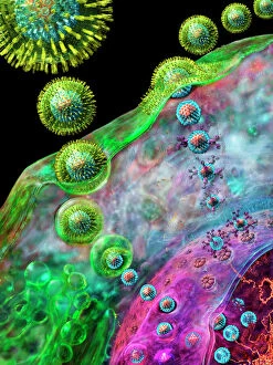
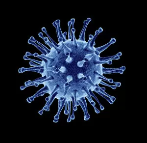
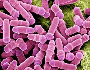
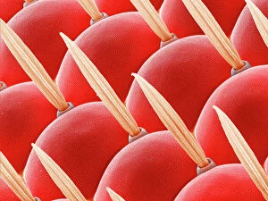
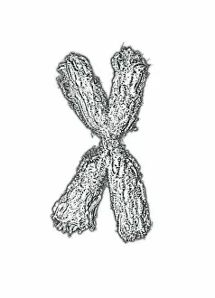
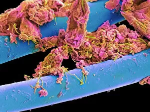
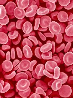
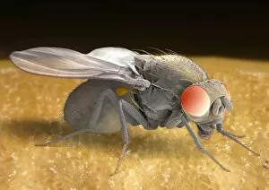
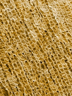
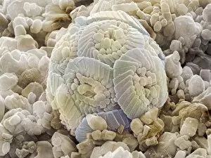
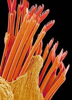
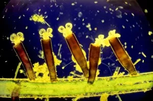
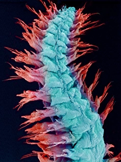
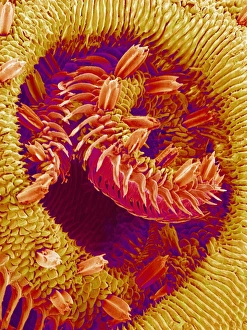
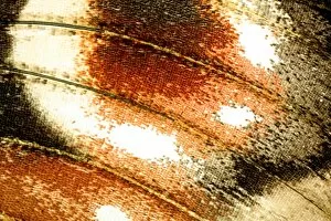
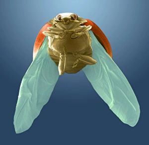
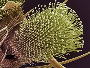
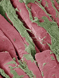
"Unveiling the Unseen: Exploring Microscopic Marvels" Delving into the intricate world of cerebellum tissue, this light micrograph reveals its complex structure and function. Witness the mesmerizing beauty of a uterus lining during menstruation, captured through a scanning electron microscope (SEM), showcasing its remarkable cellular changes. The avian flu virus, magnified under high resolution, exposes its menacing spikes that make it a formidable threat to both birds and humans alike. Discovering art in unexpected places - behold the woven fabric as you've never seen it before. SEM unveils the delicate patterns and textures hidden within this everyday material. Witness life's eternal cycle with dividing yeast cells captured through SEM, offering an up-close look at their fascinating reproduction process. A haunting image emerges as we observe an avian flu virus particle lurking in our midst; SEM captures its eerie form and reminds us of the importance of vigilance against such pathogens. Dive into the microscopic battlefield where bacteria infect macrophages – immune system warriors – revealing their struggle for survival in stunning detail through SEM imaging. Enter the captivating realm of insects with a compound eye of a fly showcased by SEM Z340 / 0698; marvel at nature's ingenious design for visual perception on such a minuscule scale. Journey deeper into brain matter with another glimpse at cerebellum tissue, now illuminated by light micrography to unravel further mysteries held within this vital organ. Explore the intricacies of our circulatory system as red blood cells come alive under SEM; witness their unique shape and learn about their crucial role in oxygen transport throughout our bodies. An unconventional view awaits as used dental floss is examined under SEM; uncovering unseen remnants left behind after oral hygiene routines take place each day.