Poster Print > Popular Themes > DNA
Poster Print : Rough endoplasmic reticulum, TEM


![]()
Poster Prints from Science Photo Library
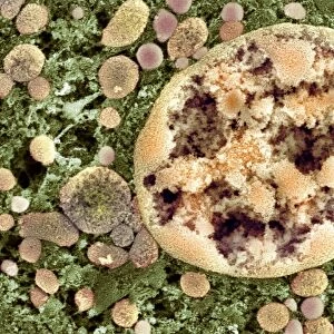
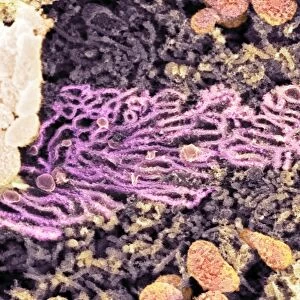
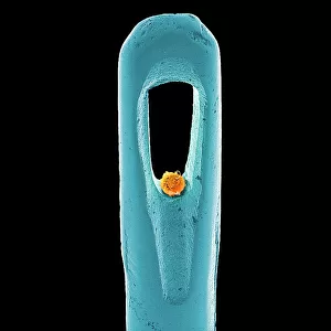
Rough endoplasmic reticulum, TEM
Rough endoplasmic reticulum, coloured transmission electron micrograph (TEM). This section shows the rough endoplasmic reticulum (ER, folds, centre), a membranous structure that occurs in cells. The cell nucleus is partially seen at left. Rough ER has ribosomes on its surface where protein synthesis occurs. The instructions for the protein synthesis come from the DNA (deoxyribonucleic acid) in the nucleus. The rounded structures may be vesicles that have broken off the rough ER to transport the proteins elsewhere in the cell. That would mean that this is the Golgi apparatus. Magnification: x20, 000 when printed 10cm wide
Science Photo Library features Science and Medical images including photos and illustrations
Media ID 6403199
© BIOMEDICAL IMAGING UNIT, SOUTHAMPTON GENERAL HOSPITAL/SCIENCE PHOTO LIBRARY
Cellular Cross Section Cytological Cytology Endoplasmic Reticulum False Colour Golgi Apparatus Histological Histology Membrane Nucleus Organelle Organelles Physiological Physiology Protein Synthesis Ribosome Ribosomes Rough Transmission Electron Microscope Vesicle Vesicles False Coloured Section Sectioned
A2 (59.4 x 42cm) Poster Print
Discover the intricacies of cellular structures with our Media Storehouse Poster Prints featuring the captivating image of "Rough Endoplasmic Reticulum, TEM" by Science Photo Library. This stunning Transmission Electron Micrograph (TEM) image showcases the rough endoplasmic reticulum (ER), a membranous structure that plays a crucial role in various cellular functions. The ER's distinctive rough surface, formed by ribosomes attached to the membrane, is vividly depicted, providing an enlightening visual representation of this essential cellular component. Bring this scientific masterpiece into your workspace or home for a daily reminder of the wonders of the cellular world.
A2 Poster (59.4 x 42cm, 23.4" x 16.5" inches) printed on 170gsm Satin Poster Paper. Securely packaged, rolled and inserted into a strong mailing tube and shipped tracked. Poster Prints are of comparable archival quality to our Photographic prints, they are simply printed on thinner Poster Paper. Whilst we only use Photographic Prints in our frames, you can frame Poster Prints if they are carefully supported to prevent sagging over time.
Poster prints are budget friendly enlarged prints in standard poster paper sizes (A0, A1, A2, A3 etc). Whilst poster paper is sometimes thinner and less durable than our other paper types, they are still ok for framing and should last many years. Our Archival Quality Photo Prints and Fine Art Paper Prints are printed on higher quality paper and the choice of which largely depends on your budget.
Estimated Image Size (if not cropped) is 53.3cm x 42cm (21" x 16.5")
Estimated Product Size is 59.4cm x 42cm (23.4" x 16.5")
These are individually made so all sizes are approximate
Artwork printed orientated as per the preview above, with landscape (horizontal) orientation to match the source image.
EDITORS COMMENTS
This print showcases the intricate structure of the rough endoplasmic reticulum (ER) in a cell. In this coloured transmission electron micrograph, we can observe the folds and membranes that make up this vital organelle. The rough ER is distinguished by the presence of ribosomes on its surface, where protein synthesis takes place. The DNA within the cell nucleus provides instructions for protein synthesis, which are then carried out by the ribosomes on the rough ER. As proteins are synthesized, they may be transported to other parts of the cell through vesicles that have broken off from the rough ER. These rounded structures visible in the image could potentially be these transport vesicles destined for another crucial cellular component –the Golgi apparatus. With a magnification of x20,000 when printed at 10cm wide, this photograph offers an extraordinary glimpse into cellular biology and physiology. It highlights not only how cells function but also emphasizes their complexity and organization at a microscopic level. This stunning image was captured using a transmission electron microscope (TEM), allowing us to explore cross-sections of cells with incredible detail. Provided by Science Photo Library, it serves as a valuable resource for researchers and enthusiasts alike who seek to delve deeper into cytology, histology, and biological sciences as a whole.
MADE IN THE UK
Safe Shipping with 30 Day Money Back Guarantee
FREE PERSONALISATION*
We are proud to offer a range of customisation features including Personalised Captions, Color Filters and Picture Zoom Tools
SECURE PAYMENTS
We happily accept a wide range of payment options so you can pay for the things you need in the way that is most convenient for you
* Options may vary by product and licensing agreement. Zoomed Pictures can be adjusted in the Basket.