Fraud Blocker Skip to main content

Nucleus Collection

"The Nucleus: Unveiling the Mysteries of Life's Command Center" Exploring the intricate world within our cerebellum tissue

Background imageNucleus Collection: Cerebellum tissue, light micrograph

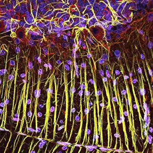
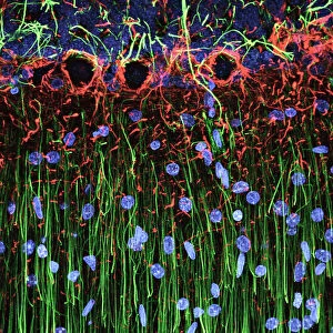
Cerebellum tissue, light micrograph
Cerebellum tissue. Confocal light micrograph of a section through the cerebellum of the brain. Purkinje cells, a type of neuron (nerve cell), are red

Background imageNucleus Collection: Hippocampus brain tissue

Hippocampus brain tissue
Hippocampus tissue. Light micrograph of a sagittal (side view) section through the hippocampus of the brain showing the nerve cells within it

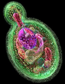
Background imageNucleus Collection: Budding yeast cell

Budding yeast cell. Computer artwork of asection through a yeast ( Candida albicans )cell that is reproducing asexually. A daughtercell (top left) is budding from the parent cell

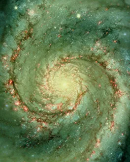
Background imageNucleus Collection: M51 whirlpool galaxy

M51 whirlpool galaxy

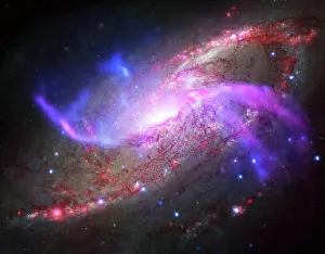
Background imageNucleus Collection: A galactic light show in spiral galaxy NGC 4258

A galactic light show in spiral galaxy NGC 4258, also known as M106, about 23 million light years away. This galaxy is famous, however

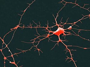
Background imageNucleus Collection: Nerve cell, SEM

Nerve cell, SEM
Nerve cell. Coloured scanning electron micrograph (SEM) of a nerve cell (neuron). Neurons are responsible for passing information around the central nervous system (CNS)

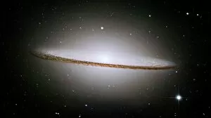
Background imageNucleus Collection: The majestic Sombrero Galaxy (Messier 104)

The majestic Sombrero Galaxy (Messier 104)

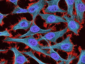
Background imageNucleus Collection: HeLa cells, light micrograph C017 / 8299

HeLa cells, light micrograph C017 / 8299
HeLa cells, multiphoton fluorescence micrograph (MFM). The cell nuclei, which contain the cells genetic information, are purple. Microtubules are blue and actin microfilaments are red

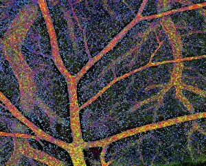
Background imageNucleus Collection: Brain tissue blood supply

Brain tissue blood supply. Light micrograph of a section through cortex tissue from a brain, showing the blood vessels (branching) that supply it

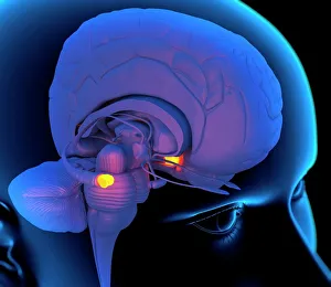
Background imageNucleus Collection: Medulla oblongata in the brain, artwork

Medulla oblongata in the brain, artwork
Hypothalamus in the brain. Computer artwork of a persons head showing the left hemisphere of the brain inside. The highlighted area shows the hypothalamus

Background imageNucleus Collection: Nuclear Fission Artwork

Nuclear Fission Artwork
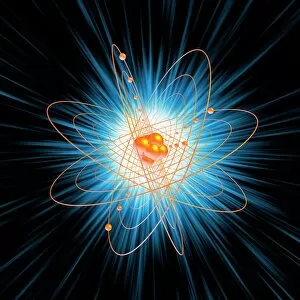
Nuclear fission. Conceptual computer artwork of an atom being split through nuclear, or atomic, fission (splitting). Electrons (orange) can be seen orbiting the nucleus (centre)

Background imageNucleus Collection: A Black Hole in a Globular Cluster

A Black Hole in a Globular Cluster
Artists View of a Black Hole in a Globular Cluster

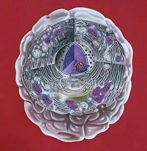
Background imageNucleus Collection: Human cell

Human cell
Illustration of a highly magnified section through a human cell. Page 8 from Human Biology, 1977

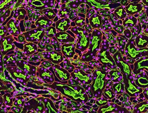
Background imageNucleus Collection: Kidney tubules in section

Kidney tubules in section
Kidney tubules. Fluorescent light micrograph of a section through kidney tissue showing numerous tubules (black/green). The tubules are seen in cross-section

Background imageNucleus Collection: Rough endoplasmic reticulum, TEM

Rough endoplasmic reticulum, TEM
Rough endoplasmic reticulum, coloured transmission electron micrograph (TEM). This section shows the rough endoplasmic reticulum (ER, folds, centre), a membranous structure that occurs in cells

Background imageNucleus Collection: Atomic structure, artwork

Atomic structure, artwork
Atomic structure. Computer artwork of electrons orbiting a central nucleus. This is a classical schematic Bohr model of an atom

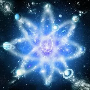
Background imageNucleus Collection: Quantised orbits of the planets

Quantised orbits of the planets
Quantum orbits of the planets. Conceptual computer artwork showing the planets of the solar system on a model of atomic orbitals

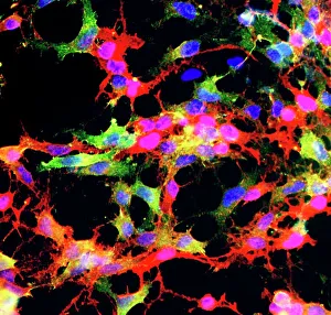
Background imageNucleus Collection: Glial stem cell culture, light micrograph

Glial stem cell culture, light micrograph
Glial stem cell culture. Fluorescent light micrograph of glial stem cells producing the protein NG2 (red) as they mature. These stem cells can differentiate into several types of glial cells

Background imageNucleus Collection: Cerebral cortex nerve cells

Cerebral cortex nerve cells. Confocal light micrograph of neurons (nerve cells, red) and glial cells (support cells, gold) from the cerebral cortex

Background imageNucleus Collection: Herpes virus replicating

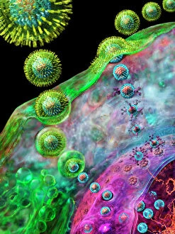
Herpes virus replicating, computer artwork.Viruses are only able to replicate in a host cell.The glycoprotein spikes in the virus envelope(green)

Background imageNucleus Collection: Cell structure

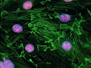
Cell structure. Confocal light micrograph of cultured endothelial cells. A fluorescent dye has been used to show the cell structure

Background imageNucleus Collection: HeLa cells, light micrograph C017 / 8298

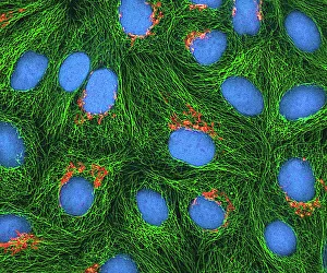
HeLa cells, light micrograph C017 / 8298
HeLa cells, multiphoton fluorescence micrograph (MFM). The cell nuclei, which contain the cells genetic information, are blue. Golgi bodies, which modify and package proteins, are orange

Background imageNucleus Collection: Mitosis, light micrograph

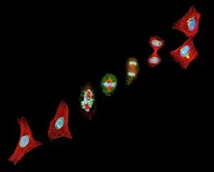
Mitosis, light micrograph
Mitosis. Confocal light micrograph of the stages of mitosis (nuclear division) and cytokinesis (cell division). During mitosis the nuclear envelope disintegrates (3rd image) and the chromosomes (blue)

Background imageNucleus Collection: Regenerating nerve cell, TEM

Regenerating nerve cell, TEM
Regenerating nerve cell. Coloured transmission electron micrograph (TEM) of a section through a nerve axon (blue) regenerating within a Schwann cell (light brown)

Background imageNucleus Collection: Trypanosome protozoan, artwork

Trypanosome protozoan, artwork
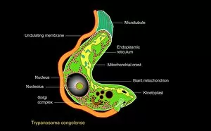
Trypanosome protozoan. Artwork of a section through a Trypanosoma congolense protozoan. This parasite is a cause of the disease nagana in cattle and other livestock in Africa

Background imageNucleus Collection: Cell types, artwork

Cell types, artwork
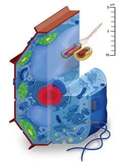
Cell types. Cutaway artwork with a scale bar (upper right, in micrometres) showing the relative sizes of eukaryotic cells (those containing a nucleus) and prokaryotic cells (those lacking a nucleus)

Background imageNucleus Collection: Basophil white blood cell

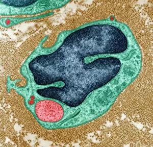
Basophil white blood cell. Coloured transmission electron micrograph (TEM) of a basophil white blood cell. Basophils are the smallest and rarest of the white blood cells

Background imageNucleus Collection: Regenerating nerve cell, TEM

Regenerating nerve cell, TEM
Regenerating nerve cell. Coloured transmission electron micrograph (TEM) of a section through a nerve axon (yellow) regenerating within a Schwann cell (blue). The Schwann cells nucleus is black

Background imageNucleus Collection: HeLa cells, light micrograph C013 / 4774

HeLa cells, light micrograph C013 / 4774
HeLa cells. Multi-photon fluorescence light micrograph of a group of cultured HeLa cells, showing the cell nuclei, which contain the cells genetic information (DNA, red)

Background imageNucleus Collection: Cerebellum tissue, light micrograph

Cerebellum tissue, light micrograph
Cerebellum tissue. Confocal light micrograph of a section through the cerebellum of the brain. Purkinje cells, a type of neuron (nerve cell), are red

Background imageNucleus Collection: James Chadwick, British physicist C017 / 7111

James Chadwick, British physicist C017 / 7111
James Chadwick (1891-1974), British physicist. Educated in Manchester, Chadwicks research under Rutherford was mainly with alpha particles (helium nuclei)

Background imageNucleus Collection: Artists concept of an astronaut falling towards a black hole in outer space

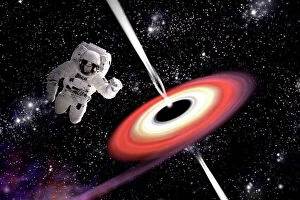
Artists concept of an astronaut falling towards a black hole in outer space. Gamma Rays erupt from the poles as a nearby nebula is pulled in

Background imageNucleus Collection: Astrocyte nerve cell

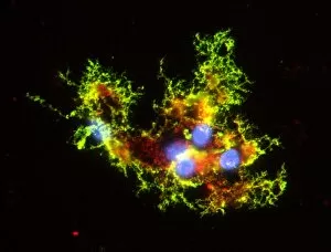
Astrocyte nerve cell. Fluorescent light micrograph of an astrocyte cell from a human brain. Intermediate filaments (IFs), part of the cells cytoskeleton, have been dyed green

Background imageNucleus Collection: Immunofluorescent LM of fibroblast cell nuclei

Immunofluorescent LM of fibroblast cell nuclei
Cell nuclei. Immunofluorescent light micrograph of cultured fibroblast cells showing nuclei with " nucleolar necklaces"

Background imageNucleus Collection: Ovarian follicle, SEM

Ovarian follicle, SEM
Ovarian follicle. Coloured scanning electron micrograph (SEM) of a fracture through a secondary follicle in the ovary. The oocyte (developing egg) is orange and its central nucleus is darker orange

Background imageNucleus Collection: Fibroblast cell, artwork

Fibroblast cell, artwork
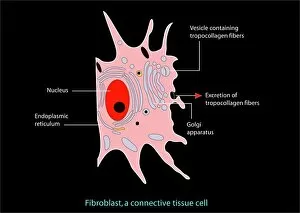
Fibroblast cell. Computer artwork of a fibroblast excreting collagen fibres (tropocollagen). Fibroblasts are cells that produce connective tissue such as collagen (tropocollagen)

Background imageNucleus Collection: Golgi apparatus, SEM

Golgi apparatus, SEM
Golgi apparatus, coloured scanning electron micrograph (SEM). Section through a liver cell showing its Golgi apparatus (grey), a membrane-bound organelle that modifies and packages proteins

Background imageNucleus Collection: Atomic structure

Atomic structure, computer artwork. Schematic representation of the structure of the atom. At the centre of the model is the nucleus, made up of a tightly packed cluster of protons and neutrons

Background imageNucleus Collection: The Whirlpool Galaxy and its companion galaxy

The Whirlpool Galaxy and its companion galaxy
The Whirlpool Galaxy (NGC 5194), and its companion galaxy (NGC 5195), in the constellation Canes Venatici

Background imageNucleus Collection: Nerve cell, TEM

Nerve cell, TEM
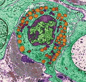
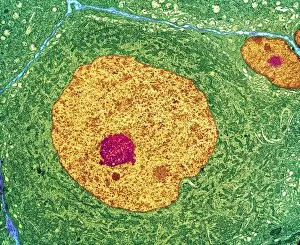
Nerve cell. Coloured transmission electron micrograph (TEM) of a nerve cell body in cross- section. The cell has a large nucleus (yellow) and inner nucleolus (red)

Background imageNucleus Collection: Oligodendrocyte nerve cells

Oligodendrocyte nerve cells. Fluorescent light micrograph of human oligodendrocyte nerve cells. Cell nuclei, which contain the cells genetic information, have been dyed blue

Background imageNucleus Collection: Purkinje nerve cell, TEM C014 / 0583

Purkinje nerve cell, TEM C014 / 0583
Purkinje nerve cell. Transmission electron micrograph (TEM) of a purkinje nerve cell (green) from the cerebellum of the brain, showing the cell body (centre) and its primary dendrite (cell process)

Background imageNucleus Collection: Milky Way galaxy

Milky Way galaxy
Milky Way. Computer artwork of our galaxy, the Milky Way, seen from above. This image is based on measurements of the galaxys stars taken in infrared, radio and other wavelengths

Background imageNucleus Collection: Cells, conceptual computer artwork

Cells, conceptual computer artwork

Background imageNucleus Collection: Lymphocyte white blood cells, artwork

Lymphocyte white blood cells, artwork
Lymphocyte white blood cells. Computer artwork of lymphocyte white blood cells, showing their nuclei (red, centre). Lymphocytes, like all white blood cells, are part of the immune system

Background imageNucleus Collection: Triangulum galaxy (M33)

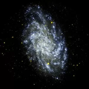
Triangulum galaxy (M33), ultraviolet image. This spiral galaxy, also known as the Pinwheel galaxy, is a member of the Local Group of galaxies

Background imageNucleus Collection: Heart muscle, confocal light micrograph

Heart muscle, confocal light micrograph
Heart muscle. Confocal light micrograph of a section through cardiac (heart) muscle. Cardiac muscle consists of branching elongated muscle cells

Background imageNucleus Collection: Model of cyclotron, particle accelerator, invented by Ernest Orlando Lawrence (1901-1958)

Model of cyclotron, particle accelerator, invented by Ernest Orlando Lawrence (1901-1958)
AIS5378122 Model of cyclotron, particle accelerator, invented by Ernest Orlando Lawrence (1901-1958) and Milton Stanley Livingston (1905-1986) in 1931 (metal object); Science Museum, London

Background imageNucleus Collection: The wonder that is electricity (litho)

The wonder that is electricity (litho)
8676245 The wonder that is electricity (litho) by English School, (20th century); Private Collection; (add.info.: The wonder that is electricity)

Background imageNucleus Collection: Animal cell structure

Animal cell structure. Artwork of a section through an animal cell. At lower centre is the nucleus, which contains the cells genetic information in the form of DNA (deoxyribonucleic acid)

Background imageNucleus Collection: Group Of Modernist Houses, Haywards Heath

Group Of Modernist Houses, Haywards Heath
A photograph of a group of modernist houses forming the nucleus of a housing estate in Haywards Heath, Sussex, designed in 1936 by architect Berthold Romanovich Lubetkin and Tecton. Date: circa 1937

Background imageNucleus Collection: Warty crab and flat rock crab

Warty crab and flat rock crab
Warty crab or yellow crab, Eriphia verrucosa 1, and flat rock crab, Percnon planissimum 2. Erifia fronte spinosa, Plagusia clavimana

Background imageNucleus Collection: Tiger cowrie, wrinkled cowrie, money cowrie, etc

Tiger cowrie, wrinkled cowrie, money cowrie, etc
Tiger cowrie, Cypraea tigris 1, wrinkled cowrie, Nucleolaria nucleus 2, money cowrie, Monetaria moneta 3, and Ellatrivia merces 4, Porcelaines

Background imageNucleus Collection: Egypt after the War, Lord Dufferin at Cairo (engraving)

Egypt after the War, Lord Dufferin at Cairo (engraving)
1061236 Egypt after the War, Lord Dufferin at Cairo (engraving) by English School, (19th century); Private Collection; (add.info.: Egypt after the War, Lord Dufferin at Cairo)

Background imageNucleus Collection: The War in South Africa (litho)

The War in South Africa (litho)
1599684 The War in South Africa (litho) by Stewart, Frank Algenon (1877-1945); Private Collection; (add.info.: The War in South Africa. Illustration for The Illustrated London News, 26 May 1900)

Background imageNucleus Collection: EyeUbiquitous_20104516

EyeUbiquitous_20104516
Food, Vegetables, Onions, Three ripe harvested onion bulbs or Allium against a white background with one sliced open to show structure of layers

Background imageNucleus Collection: Ian Carr, Nucleus, Ronnie Scotts, Soho London, May 1991. Creator: Brian O Connor

Ian Carr, Nucleus, Ronnie Scotts, Soho London, May 1991. Creator: Brian O Connor
Ian Carr, Nucleus, Ronnie Scott s, Soho, London, May 1991

Background imageNucleus Collection: Illustration of cell nucleus containing cell cytoplasm, mitochondria units, DNA, and chromosome

Illustration of cell nucleus containing cell cytoplasm, mitochondria units, DNA, and chromosome

Background imageNucleus Collection: Illustration of atom with nucleus of protons and neurons, based on the Bohr model

Illustration of atom with nucleus of protons and neurons, based on the Bohr model

Background imageNucleus Collection: Microscopic view of animal cell

Microscopic view of animal cell

Background imageNucleus Collection: Oxygen atomic structure, artwork

Oxygen atomic structure, artwork
Oxygen atomic structure. Computer artwork showing the structure of an oxygen atom. Each oxygen atom contains 8 electrons (blue) orbiting the atomic nucleus (centre)

Background imageNucleus Collection: Helium, atomic model

Helium, atomic model
Heium, atomic model. Helium has two neutrons (white) and two protons (pink) in its nucleus (centre). The atom also has two electron (blue) orbiting the nucleus

Background imageNucleus Collection: Animal cell anatomy, diagram

Animal cell anatomy, diagram
Animal cell anatomy. Diagram showing the internal and external anatomy of an animal cell

Background imageNucleus Collection: A row of taxi cabs waiting to take Automobile Association offiers

A row of taxi cabs waiting to take Automobile Association offiers and patrols to the White City to form the nucleus of a new cyclist battalion, the Army Cyclist Corps, at the start of WWI

Background imageNucleus Collection: Cross section illustration of human cell

Cross section illustration of human cell

Background imageNucleus Collection: Illustration of Rutherford model of the atom, consisting of nucleus and negatively charged electrons

Illustration of Rutherford model of the atom, consisting of nucleus and negatively charged electrons

Background imageNucleus Collection: Illustration of neutron hitting Uranium-235 nucleus, nucleus becoming unstable and splitting

Illustration of neutron hitting Uranium-235 nucleus, nucleus becoming unstable and splitting, releasing energy and neutrons (nuclear fission)

Background imageNucleus Collection: Microscopic view of cell

Microscopic view of cell

Background imageNucleus Collection: Retina with macular degeneration

Retina with macular degeneration

Background imageNucleus Collection: Cluster of HIV virus

Cluster of HIV virus. HIV is the human immunodeficiency virus that can lead to acquired immune deficiency syndrom, or AIDS

Background imageNucleus Collection: Arp 273 interacting galaxies in Andromeda

Arp 273 interacting galaxies in Andromeda. The larger of the two galaxies, UGC 1810, shows a large spiral galaxy with a disk that is distorted into a rose-like shape by the gravitational tidal pull

Background imageNucleus Collection: Antennae colliding galaxies C017 / 3745

Antennae colliding galaxies C017 / 3745
Antennae colliding galaxies, combined optical image. The Antennae (NGC 4038 and NGC 4039) are formed of two galaxies colliding due to mutual gravitational attraction

Background imageNucleus Collection: Comet over trees, artwork C015 / 0777

Comet over trees, artwork C015 / 0777
Comet over trees, artwork. Comets are bodies of ice and dust that enter the inner solar system from the outer solar system

Background imageNucleus Collection: Macrophage white blood cells, TEM

Macrophage white blood cells, TEM
Macrophage white blood cells. Transmission electron micrograph (TEM) of a section through two macrophages located within connective tissue, showing the nuclei and numerous lysosomes (dark spots)

Background imageNucleus Collection: Pancreatic acinar cell

Pancreatic acinar cell. Transmission electron micrograph (TEM) of a section through an enzyme-secreting acinar cell in the human pancreas, showing part of the nucleus (round, far left)

Background imageNucleus Collection: Smooth muscle tissue, TEM

Smooth muscle tissue, TEM
Smooth muscle tissue. Transmission electron micrograph (TEM) of a longitudinal section through smooth muscle cells. Smooth muscle cells are spindle-shaped and often closely packed together

Background imageNucleus Collection: Cartilage cell, TEM C014 / 1433

Cartilage cell, TEM C014 / 1433
Cartilage cell. Transmission electron micrograph (TEM) of a section through a chondrocyte cell from hyaline cartilage of the trachea (windpipe)

Background imageNucleus Collection: Cartilage cell, TEM C014 / 1434

Cartilage cell, TEM C014 / 1434
Cartilage cell. Transmission electron micrograph (TEM) of a section through a chondrocyte cell from hyaline cartilage of the trachea (windpipe)

Background imageNucleus Collection: Nerve cell growth

Nerve cell growth. Light micrograph of nerve cells (neurons) with immunofluorescent staining. These cells have been grown in culture

Background imageNucleus Collection: Beryllium, atomic model

Beryllium, atomic model. Beryllium has five neutrons (white) and four protons (pink) in its nucleus (centre). The atom also has four electron (blue) orbiting the nucleus

Background imageNucleus Collection: Boron, atomic model

Boron, atomic model. Boron has six neutrons (white) and five protons (pink) in its nucleus (centre). The atom also has five electron (blue) orbiting the nucleus

Background imageNucleus Collection: English Cathedrals - Gloucester - the Nave

English Cathedrals - Gloucester - the Nave - Ornamental stone arches between the columns. Part of Box 144 Boswell Collection - English Cathedrals

Background imageNucleus Collection: Spain. Citadel of Roses (Rhode). Roman ruins

Spain. Citadel of Roses (Rhode). Roman ruins
Spain. Citadel of Roses (Rhode). Excavations of a urban nucleus of the Low Roman Empire. 3rd-2nd centuries BC

Background imageNucleus Collection: Pre-romanesque Church of Saint Peter. Terrassa. Spain

Pre-romanesque Church of Saint Peter. Terrassa. Spain
Pre-romanesque Church of Saint Peter. View of the trefoil apse. 9th-10th centuries. Terrassa. Catalonia. Spain


All Professionally Made to Order for Quick Shipping

"The Nucleus: Unveiling the Mysteries of Life's Command Center" Exploring the intricate world within our cerebellum tissue, where the nucleus orchestrates every movement and balance. A mesmerizing light micrograph reveals the nucleus at the heart of M51 Whirlpool Galaxy, a cosmic dance guided by its gravitational pull. Witnessing life's beginning through a budding yeast cell, as its nucleus holds the blueprint for growth and reproduction. Delving into the secrets of communication with a nerve cell captured in stunning detail through SEM imaging, showcasing its complex nucleus. Behold the majestic Sombrero Galaxy (Messier 104), where billions of stars revolve around their central nucleus like celestial dancers in perfect harmony. The hippocampus brain tissue unveils memories etched within each neuron's nucleus, shaping who we are and what we remember. Peering into HeLa cells under a light microscope (C017/8299), revealing their remarkable nuclei that have revolutionized medical research. An artistic representation takes us deep into Medulla Oblongata's core—the vital center controlling essential bodily functions—where nuclei reign supreme. Captivating artwork depicts nuclear fission—a powerful force harnessed to generate energy while reminding us of its immense potential and responsibility. Marvel at NGC 4258 spiral galaxy's galactic light show, where vibrant colors illuminate star clusters revolving around their radiant nuclear core. Human cells come alive under scrutiny as their nuclei hold genetic information that shapes our unique characteristics and existence itself. Exploring kidney tubules in section unravels an intricate network governed by countless tiny nuclei working harmoniously to maintain our body’s equilibrium. Intricate yet awe-inspiring, these glimpses into various realms remind us that no matter how vast or minuscule, from galaxies to microscopic cells - all bow to the commanding presence of the nucleus.

© Copyright Media Storehouse, All Rights Reserved 2003 - 2024
Facebook     Pinterest     Card Payments     Reviews IO