Fraud Blocker Skip to main content

Endoplasmic Reticulum Collection

The endoplasmic reticulum (ER) is a complex network of membranes found in eukaryotic cells and can be divided into two main types

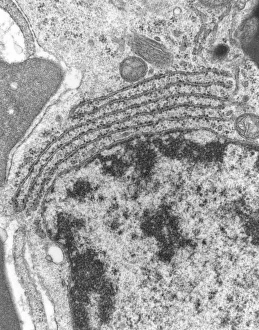
Background imageEndoplasmic Reticulum Collection: Rough endoplasmic reticulum, TEM

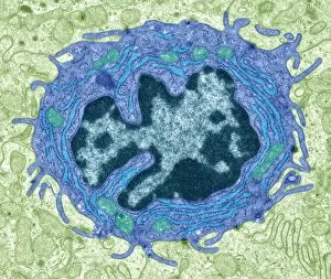
Rough endoplasmic reticulum, TEM
Rough endoplasmic reticulum, coloured transmission electron micrograph (TEM). This section shows the rough endoplasmic reticulum (ER, folds, centre), a membranous structure that occurs in cells

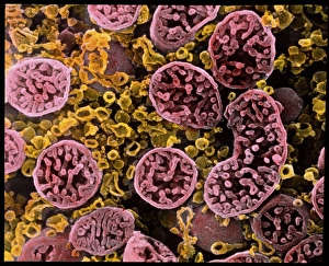
Background imageEndoplasmic Reticulum Collection: Coloured SEM of mitochondria in ovarian cells

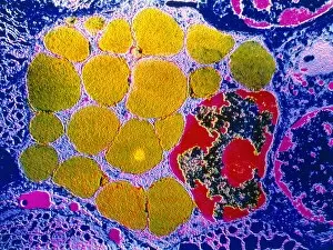
Coloured SEM of mitochondria in ovarian cells
Mitochondria. Coloured Scanning Electron Micro- graph (SEM) of mitochondria and smooth endoplasmic reticulum in an ovarian granulosa- lutein cell

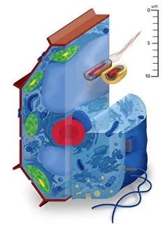
Background imageEndoplasmic Reticulum Collection: Cell types, artwork

Cell types, artwork
Cell types. Cutaway artwork with a scale bar (upper right, in micrometres) showing the relative sizes of eukaryotic cells (those containing a nucleus) and prokaryotic cells (those lacking a nucleus)

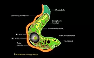
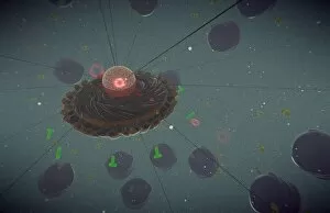
Background imageEndoplasmic Reticulum Collection: Trypanosome protozoan, artwork

Trypanosome protozoan, artwork
Trypanosome protozoan. Artwork of a section through a Trypanosoma congolense protozoan. This parasite is a cause of the disease nagana in cattle and other livestock in Africa

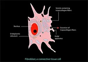
Background imageEndoplasmic Reticulum Collection: Fibroblast cell, artwork

Fibroblast cell, artwork
Fibroblast cell. Computer artwork of a fibroblast excreting collagen fibres (tropocollagen). Fibroblasts are cells that produce connective tissue such as collagen (tropocollagen)

Background imageEndoplasmic Reticulum Collection: Purkinje nerve cell, TEM C014 / 0583

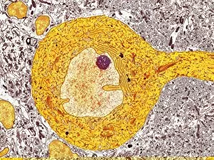
Purkinje nerve cell, TEM C014 / 0583
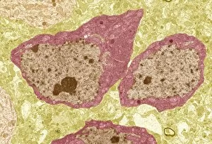
Purkinje nerve cell. Transmission electron micrograph (TEM) of a purkinje nerve cell (green) from the cerebellum of the brain, showing the cell body (centre) and its primary dendrite (cell process)

Background imageEndoplasmic Reticulum Collection: Animal cell structure

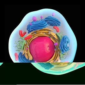
Animal cell structure. Artwork of a section through an animal cell. At lower centre is the nucleus, which contains the cells genetic information in the form of DNA (deoxyribonucleic acid)

Background imageEndoplasmic Reticulum Collection: Microscopic view of animal cell

Microscopic view of animal cell

Background imageEndoplasmic Reticulum Collection: Animal cell anatomy, diagram

Animal cell anatomy, diagram
Animal cell anatomy. Diagram showing the internal and external anatomy of an animal cell

Background imageEndoplasmic Reticulum Collection: Hepatocyte liver cell, TEM

Hepatocyte liver cell, TEM
Hepatocyte liver cell. Transmission electron micrograph (TEM) of a section through an hepatocyte liver cell, showing rough and smooth endoplasmic reticulum (ER, dark lines)

Background imageEndoplasmic Reticulum Collection: Liver tissue, TEM

Liver tissue, TEM
Liver tissue. Transmission electron micrograph (TEM) of a section through the liver, showing part of a radial cord of hepatocyte liver cells (dark) and the vascular sinusoids (white)

Background imageEndoplasmic Reticulum Collection: Skin cancer cell, SEM C016 / 4764

Skin cancer cell, SEM C016 / 4764
Skin cancer cell. Coloured ion-abrasion scanning electron micrograph (IA-SEM) of a melanoma cell. The large dark sphere is the cell nucleus, which contains the cells genetic information

Background imageEndoplasmic Reticulum Collection: TEM of endoplasmic reticulum in mammalian cell

TEM of endoplasmic reticulum in mammalian cell
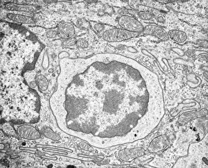
Rough endoplasmic reticulum. Transmission Electron Micrograph (TEM) of a section through a mammalian cell revealing rough endoplasmic reticulum (ER)

Background imageEndoplasmic Reticulum Collection: Cross section illustration of human cell

Cross section illustration of human cell

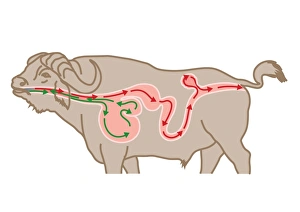
Background imageEndoplasmic Reticulum Collection: Digital cross section illustration of four chambers in stomach of ruminant, showing rumen, reticulum

Digital cross section illustration of four chambers in stomach of ruminant, showing rumen, reticulum

Background imageEndoplasmic Reticulum Collection: Conceptual image of a plant cell and its components

Conceptual image of a plant cell and its components

Background imageEndoplasmic Reticulum Collection: 3D structure of melanoma cell

3D structure of melanoma cell
3D structure of a melanoma cell derived by ion abrasion scanning electron microscopy

Background imageEndoplasmic Reticulum Collection: The interior of an eukaryotic cell

The interior of an eukaryotic cell. This is the most common generic type of cell and its present in all mammals. In the center is the nucleus (the perforated sphere)

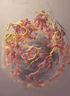
Background imageEndoplasmic Reticulum Collection: Conceptual image of endoplasmic reticulum around a cell nucleus

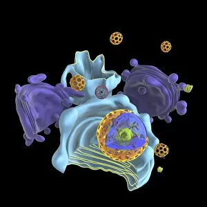
Conceptual image of endoplasmic reticulum around a cell nucleus. Endoplasmic reticulum is an organelle that forms a continuous membrane system of flattened sacs within the cytoplasm of a eukaryotic

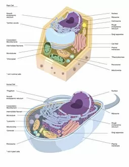
Background imageEndoplasmic Reticulum Collection: Comparative illustration of plant and animal cell anatomy (with labels)

Comparative illustration of plant and animal cell anatomy (with labels)

Background imageEndoplasmic Reticulum Collection: Model of animal cell, including cell nucleus, golgi body, lysosomes, centrioles, mitochondria

Model of animal cell, including cell nucleus, golgi body, lysosomes, centrioles, mitochondria, endoplasmic reticulum, ribosomes, cytoplasm, vesicles, thin plasma membrane

Background imageEndoplasmic Reticulum Collection: Intestinal gland cell, TEM C014 / 1441

Intestinal gland cell, TEM C014 / 1441
Intestinal gland cell. Transmission electron micrograph (TEM) of a section through an undifferentiated cell from the intestinal tract, showing extensive rough endoplasmic reticulum (ER)

Background imageEndoplasmic Reticulum Collection: Intestinal gland cell, TEM C014 / 1440

Intestinal gland cell, TEM C014 / 1440
Intestinal gland cell. Transmission electron micrograph (TEM) of a section through an undifferentiated cell from the intestinal tract, showing extensive rough endoplasmic reticulum (ER)

Background imageEndoplasmic Reticulum Collection: Animal cell, artwork F006 / 9019

Animal cell, artwork F006 / 9019
Animal cell, cut-away computer artwork

Background imageEndoplasmic Reticulum Collection: Plant cell, artwork F006 / 9876

Plant cell, artwork F006 / 9876
Plant cell, computer artwork

Background imageEndoplasmic Reticulum Collection: Endoplasmic reticulum chaperone protein F006 / 9623

Endoplasmic reticulum chaperone protein F006 / 9623
Endoplasmic reticulum chaperone protein. Molecular model of the endoplasmic reticulum chaperone protein GRP94. This protein is essential for the maturation of cell-surface display proteins

Background imageEndoplasmic Reticulum Collection: Activated plasma cell, TEM

Activated plasma cell, TEM
Activated plasma cell, coloured transmission electron micrograph (TEM). Plasma cells are derived from stimulated B lymphocytes (white blood cells)

Background imageEndoplasmic Reticulum Collection: Golgi membranes, TEM

Golgi membranes, TEM
Golgi membranes. Transmission electron micrograph (TEM) of a section through a cell, showing the membranes (dark lines) of the Golgi apparatus

Background imageEndoplasmic Reticulum Collection: Lymphocyte white blood cell, TEM

Lymphocyte white blood cell, TEM
Lymphocyte white blood cell. Transmission electron micrograph (TEM) of a section through lymphocyte, showing the nucleus (large, round, centre), endoplasmic reticulum (ER)

Background imageEndoplasmic Reticulum Collection: Eukaryote cell, artwork C016 / 6260

Eukaryote cell, artwork C016 / 6260
Eukaryote cell. Computer artwork showing the internal structure of a typical eukaryotic cell. Eukaryotes are organisms whose cells contain a membrane-bound nucleus (karyon, orange)

Background imageEndoplasmic Reticulum Collection: Animal cell, illustration C018 / 0734

Animal cell, illustration C018 / 0734
Animal cell. Illustration showing the organelles present in an animal cell. At lower centre is the nucleus, which contains the cells genetic information in the form of DNA (deoxyribonucleic acid)

Background imageEndoplasmic Reticulum Collection: Intestinal gland cell, TEM C014 / 1442

Intestinal gland cell, TEM C014 / 1442
Intestinal gland cell. Transmission electron micrograph (TEM) of a section through an undifferentiated cell from the intestinal tract, showing extensive rough endoplasmic reticulum (ER)

Background imageEndoplasmic Reticulum Collection: Monocyte white blood cell, TEM

Monocyte white blood cell, TEM
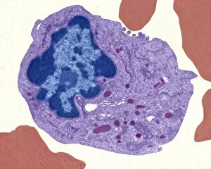
Monocyte white blood cell. Transmission electron micrograph (TEM) of a section through a monocyte white blood cell, showing the cell nucleus (blue)

Background imageEndoplasmic Reticulum Collection: Peroxiredoxin 4 antioxidant enzyme C015 / 7022

Peroxiredoxin 4 antioxidant enzyme C015 / 7022
Peroxiredoxin 4 antioxidant enzyme, molecular model. This enzyme, also called peroxiredoxin IV (PrxIV), plays a catalytic role in cell metabolism on the endoplasmic reticulum

Background imageEndoplasmic Reticulum Collection: Animal cell organelles, artwork C016 / 0619

Animal cell organelles, artwork C016 / 0619
Computer artwork showing the organelles in a eukaryotic cell. This is an animal cell. Structures include the nucleus (centre, red) which has a membrane with nuclear pores

Background imageEndoplasmic Reticulum Collection: Animal cell organelles, artwork C016 / 0621

Animal cell organelles, artwork C016 / 0621
Computer artwork showing the organelles in a eukaryotic cell. This is an animal cell. Structures include the nucleus (centre, red) which has a membrane with nuclear pores

Background imageEndoplasmic Reticulum Collection: Animal cell organelles, artwork C016 / 0617

Animal cell organelles, artwork C016 / 0617
Computer artwork showing the organelles in a eukaryotic cell. This is an animal cell. Structures include the nucleus (centre, red) which has a membrane with nuclear pores

Background imageEndoplasmic Reticulum Collection: Animal cell organelles, artwork C016 / 0611

Animal cell organelles, artwork C016 / 0611
Computer artwork showing the organelles in a eukaryotic cell. This is an animal cell. Structures include the nucleus (centre, red) which has a membrane with nuclear pores

Background imageEndoplasmic Reticulum Collection: Animal cell organelles, artwork C016 / 0612

Animal cell organelles, artwork C016 / 0612
Computer artwork showing the organelles in a eukaryotic cell. This is an animal cell. Structures include the nucleus (centre, orange) which has a membrane with nuclear pores

Background imageEndoplasmic Reticulum Collection: Animal cell organelles, artwork C016 / 0605

Animal cell organelles, artwork C016 / 0605
Computer artwork showing the organelles in a eukaryotic cell. This is an animal cell. Structures include the nucleus (centre, red) which has a membrane with nuclear pores

Background imageEndoplasmic Reticulum Collection: Animal cell organelles, artwork C016 / 0610

Animal cell organelles, artwork C016 / 0610
Computer artwork showing the organelles in a eukaryotic cell. This is an animal cell. Structures include the nucleus (centre, orange) which has a membrane with nuclear pores

Background imageEndoplasmic Reticulum Collection: Animal cell organelles, artwork C016 / 0604

Animal cell organelles, artwork C016 / 0604
Computer artwork showing the organelles in a eukaryotic cell. This is an animal cell. Structures include the nucleus (centre, red) which has a membrane with nuclear pores

Background imageEndoplasmic Reticulum Collection: Animal cell organelles, artwork C016 / 0606

Animal cell organelles, artwork C016 / 0606
Computer artwork showing the organelles in a eukaryotic cell. This is an animal cell. Structures include the nucleus (centre, orange) which has a membrane with nuclear pores

Background imageEndoplasmic Reticulum Collection: Purkinje nerve cell, TEM C014 / 0582

Purkinje nerve cell, TEM C014 / 0582
Purkinje nerve cell. Transmission electron micrograph (TEM) of a purkinje nerve cell (bright yellow, centre) from the cerebellum of the brain

Background imageEndoplasmic Reticulum Collection: Animal cell, artwork C013 / 9985

Animal cell, artwork C013 / 9985
Animal cell. Computer artwork showing the cell organelles found inside a typical animal cell. The nucleus (large round) can be seen at centre

Background imageEndoplasmic Reticulum Collection: Animal cell, artwork C013 / 9984

Animal cell, artwork C013 / 9984
Animal cell. Computer artwork showing the cell organelles found inside a typical animal cell. The nucleus (large round) can be seen at centre

Background imageEndoplasmic Reticulum Collection: Brain cells, TEM C013 / 4801

Brain cells, TEM C013 / 4801
Brain cells. Transmission electron micrograph (TEM) of a section through oligodendrocytes (dark) in human brain tissue, showing free ribosomes (dark green dots), golgi apparatus (curved brown lines)

Background imageEndoplasmic Reticulum Collection: Brain cells, TEM C013 / 4800

Brain cells, TEM C013 / 4800
Brain cells. Transmission electron micrograph (TEM) of a section through oligodendrocytes in human brain tissue, showing free ribosomes (dark pink dots), golgi apparatus (curved brown lines)


All Professionally Made to Order for Quick Shipping

The endoplasmic reticulum (ER) is a complex network of membranes found in eukaryotic cells and can be divided into two main types: rough endoplasmic reticulum (RER) and smooth endoplasmic reticulum (SER). The RER is studded with ribosomes, giving it a rough appearance under the transmission electron microscope (TEM). In contrast, the SER lacks ribosomes and appears smooth. One fascinating aspect of the ER is its involvement in various cellular processes. For instance, TEM images reveal coloured scanning electron microscopy (SEM) of mitochondria in ovarian cells, highlighting their close association with the ER. Different cell types also showcase unique artwork representations, such as trypanosome protozoan or Purkinje nerve cell. Artwork depicting fibroblast cells emphasizes their role in tissue repair and collagen production. Understanding animal cell structure becomes easier when observing hepatocyte liver cells through TEM or skin cancer cells via SEM. Cross-section illustrations provide insights into human cell organization while digital cross-sections illustrate the four chambers of a ruminant's stomach, including rumen and reticulum compartments. Lastly, conceptual images help visualize plant cell components like chloroplasts and vacuoles. Exploring the world offers a glimpse into cellular intricacies across different organisms and tissues.