Postcard > Popular Themes > DNA
Postcard : Human chromosomes, SEM C013 / 5002


![]()
Cards from Science Photo Library
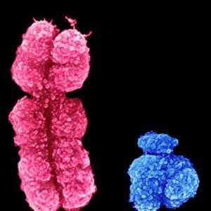
Human chromosomes, SEM C013 / 5002
Human chromosomes. Coloured scanning electron micrograph (SEM) of human chromosomes prepared with the harlequin staining technique. Chromosomes are a packaged form of a cells genetic material DNA (deoxyribonucleic acid). The DNA condenses into chromosomes during cell replication. Each chromosome consists of two identical strands (chromatids), aligned parallel to each other and joined at an area called the centromere. The staining shows the formation sequence, with the second chromatid (white) of each pair formed as a direct copy of the first chromatid (purple). Structural changes are also seen
Science Photo Library features Science and Medical images including photos and illustrations
Media ID 9195509
© SCIENCE PHOTO LIBRARY
Cell Biology Cellular Centromere Chromatid Chromatids Chromosomal Chromosome Chromosomes Colored False Color False Colored False Colour Genetic Genetic Material Staining Telomere Telomeres Biochemical Biochemistry Cutouts False Coloured Genetics
Postcards (8 pack of A6)
Set of 8, A6 Postcards, featuring the same image on all cards in a set. Printed on 350gsm premium white satin card, the back of the postcard includes space to write messages and an area for the address and stamp. Size of each postcard is 15cm x 10.6cm.
Photo postcards are a great way to stay in touch with family and friends.
Estimated Product Size is 10.6cm x 15cm (4.2" x 5.9")
These are individually made so all sizes are approximate
Artwork printed orientated as per the preview above, with landscape (horizontal) or portrait (vertical) orientation to match the source image.
FEATURES IN THESE COLLECTIONS
> Popular Themes
> DNA
EDITORS COMMENTS
This print showcases the intricate beauty of human chromosomes, captured through a scanning electron microscope (SEM) using the harlequin staining technique. The image reveals the packaged form of a cell's genetic material, DNA (deoxyribonucleic acid), which condenses into chromosomes during cell replication. Each chromosome in this mesmerizing display consists of two identical strands called chromatids, aligned parallel to each other and joined at an area known as the centromere. The stunning false coloration highlights the formation sequence, with the second chromatid appearing white as it is formed as a direct copy of its purple counterpart. Beyond their aesthetic appeal, these chromosomes hold immense significance in understanding our genetic makeup. They serve as carriers of vital information that determines various aspects of our biology and health. Structural changes within these chromosomes are also visible in this image, providing valuable insights into cellular processes and genetic mutations. The black background enhances the visual impact while emphasizing the vibrant colors that represent different components within these chromosomal structures. This remarkable photograph serves as a testament to both scientific discovery and artistic expression, showcasing how even microscopic elements can possess astonishing beauty when observed through advanced imaging techniques like SEM.
MADE IN THE UK
Safe Shipping with 30 Day Money Back Guarantee
FREE PERSONALISATION*
We are proud to offer a range of customisation features including Personalised Captions, Color Filters and Picture Zoom Tools
SECURE PAYMENTS
We happily accept a wide range of payment options so you can pay for the things you need in the way that is most convenient for you
* Options may vary by product and licensing agreement. Zoomed Pictures can be adjusted in the Basket.