Fraud Blocker Skip to main content

Chromosome Collection

"Unlocking the Mysteries of Life: Exploring the World of Chromosomes" Witness the intricate dance of life as cells divide through mitosis

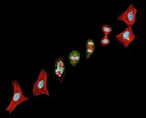
Background imageChromosome Collection: Mitosis, light micrograph

Mitosis, light micrograph
Mitosis. Confocal light micrograph of the stages of mitosis (nuclear division) and cytokinesis (cell division). During mitosis the nuclear envelope disintegrates (3rd image) and the chromosomes (blue)

Background imageChromosome Collection: Cell division, fluorescent micrograph

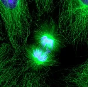
Cell division, fluorescent micrograph
Cell division. Immunofluorescent light micrograph of a human epithelial cell (centre) during the late anaphase stage of mitosis

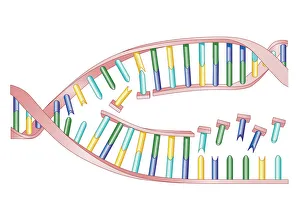
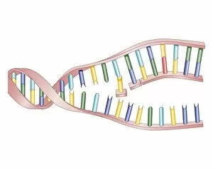
Background imageChromosome Collection: Biomedical illustration of DNA Replication as bases attach to strand with two newly formed strands

Biomedical illustration of DNA Replication as bases attach to strand with two newly formed strands twist eventually producing two new identical double DNA strands

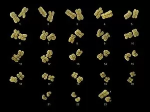
Background imageChromosome Collection: Full set of male chromosomes, SEM

Full set of male chromosomes, SEM
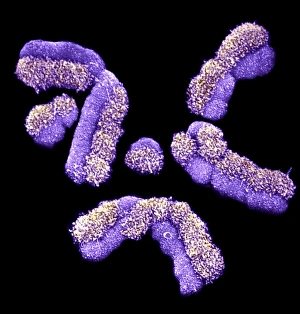
Set of human chromosomes, coloured scanning election micrograph (SEM). Chromosomes are a packaged form of the genetic material deoxyribonucleic acid (DNA)

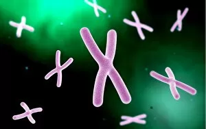
Background imageChromosome Collection: Chromosome

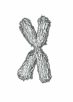
Chromosome. Computer artwork of a condensed chromosome. Chromosomes, which consist of two identical chromatids joined at a centromere (centre)

Background imageChromosome Collection: Dividing cells

Dividing cells. Computer artwork of a spiral of cells during mitosis (nuclear division). During mitosis two daughter nuclei are formed from one parent nucleus

Background imageChromosome Collection: Cell division, fluorescent micrograph

Cell division, fluorescent micrograph
Cell division. Immunofluorescent light micrograph of a human epithelial cell (centre) during the interphase stage of mitosis

Background imageChromosome Collection: Illustration of chromosome structure

Illustration of chromosome structure

Background imageChromosome Collection: Cross section biomedical illustration of protein synthesis and ribosome

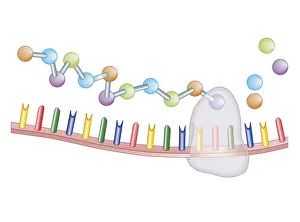
Cross section biomedical illustration of protein synthesis and ribosome

Background imageChromosome Collection: Astrocyte nerve cell

Astrocyte nerve cell. Fluorescent light micrograph of an astrocyte cell from a human brain. Intermediate filaments (IFs), part of the cells cytoskeleton, have been dyed green

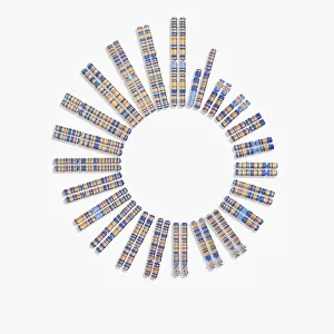
Background imageChromosome Collection: Human chromosomes, SEM C013 / 5002

Human chromosomes, SEM C013 / 5002
Human chromosomes. Coloured scanning electron micrograph (SEM) of human chromosomes prepared with the harlequin staining technique

Background imageChromosome Collection: Illustration showing DNA replication

Illustration showing DNA replication
Beginnings, Biology, Biomedical Illustration, Chromosome, Close-Up, Complexity, Connection, Development, Dna, Fragility, Genetic Research, Growth, Healthcare and Medicine, Helix, Identity

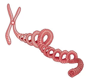
Background imageChromosome Collection: Illustration of spiral of DNA attached to chromosome

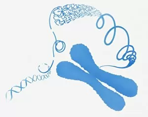
Illustration of spiral of DNA attached to chromosome
Beginnings, Biomedical Illustration, Chromosome, Close-Up, Complexity, Discovery, Dna, Full Length, Genetic Research, Healthcare and Medicine, Identity, Individuality, New Life, No People, Science

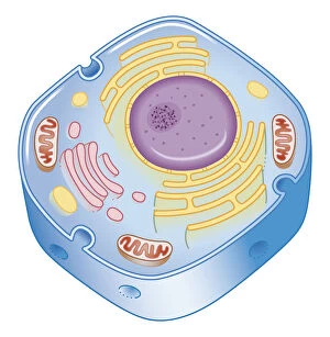
Background imageChromosome Collection: Illustration of cell nucleus containing cell cytoplasm, mitochondria units, DNA, and chromosome

Illustration of cell nucleus containing cell cytoplasm, mitochondria units, DNA, and chromosome

Background imageChromosome Collection: Plant cell mitosis, light micrograph

Plant cell mitosis, light micrograph
Plant cell mitosis. Light micrograph of root tip cells from an onion (Allium sp.) undergoing cell division (mitosis). From top left to bottom right

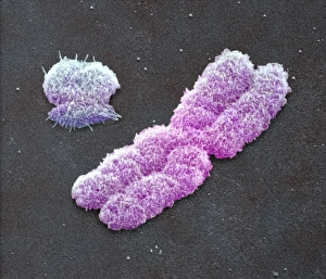
Background imageChromosome Collection: Male sex chromosomes, SEM

Male sex chromosomes, SEM
Male sex chromosomes. Coloured scanning electron micrograph (SEM) of human X (centre) and Y (upper left) sex chromosomes. Each chromosome has replic- ated to form two identical strands (chromatids)

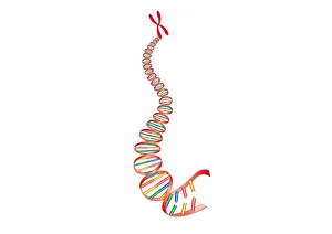
Background imageChromosome Collection: Digital illustration of animal DNA and chromosome

Digital illustration of animal DNA and chromosome

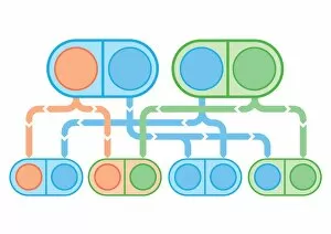
Background imageChromosome Collection: Digital illustration of X-linked recessive inheritance

Digital illustration of X-linked recessive inheritance

Background imageChromosome Collection: Digital illustration showing temporary cell damage to genes on chromosomes caused by an attacking

Digital illustration showing temporary cell damage to genes on chromosomes caused by an attacking carcinogen

Background imageChromosome Collection: Digital illustration showing human cell division with chromosome making exact copy of itself and div

Digital illustration showing human cell division with chromosome making exact copy of itself and div

Background imageChromosome Collection: Digital illustration of 22 pairs of chromosomes plus one sex pair

Digital illustration of 22 pairs of chromosomes plus one sex pair

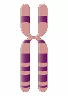
Background imageChromosome Collection: Illustration of human X chromosome

Illustration of human X chromosome

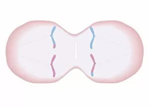
Background imageChromosome Collection: Cross section biomedical illustration of mitosis where nucleus membrane form around each set of

Cross section biomedical illustration of mitosis where nucleus membrane form around each set of chromosomes and the cell begins to divide in two

Background imageChromosome Collection: Cross section biomedical illustration of mitosis with new cells form

Cross section biomedical illustration of mitosis with new cells form, each having a central nucleus containing identical set of chromosomes

Background imageChromosome Collection: Cross section biomedical illustration of meiosis with duplicated chromosomes lined up

Cross section biomedical illustration of meiosis with duplicated chromosomes lined up and more threads attach, pulling the duplicated chromosomes apart to form two single chromosomes

Background imageChromosome Collection: Biomedical illustration of X Chromosome

Biomedical illustration of X Chromosome

Background imageChromosome Collection: Biomedical illustration of DNA Replication

Biomedical illustration of DNA Replication

Background imageChromosome Collection: Biomedical illustration of protein synthesis within DNA

Biomedical illustration of protein synthesis within DNA

Background imageChromosome Collection: Conceptual image of a telomere

Conceptual image of a telomere. A telomere is a region of the DNA sequence at the end of a chromosome. Their function is to protect the ends of the chromosome from degradating

Background imageChromosome Collection: Cell nucleus with chromosome

Cell nucleus with chromosome. The cell nucleus helps control eating, movement, and reproduction

Background imageChromosome Collection: Microscopic view of telomeres highlighted at the tips of chromosome

Microscopic view of telomeres highlighted at the tips of chromosome. A telomere is a region of the DNA sequence at the end of a chromosome

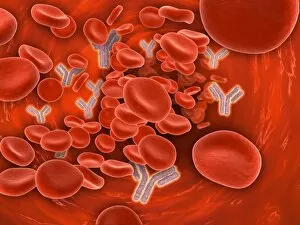
Background imageChromosome Collection: Conceptual image of chromosomes inside the blood stream

Conceptual image of chromosomes inside the blood stream

Background imageChromosome Collection: Conceptual image of a telomere showing DNA structure

Conceptual image of a telomere showing DNA structure. A telomere is a region of the DNA sequence at the end of a chromosome. Their function is to protect the ends of the chromosome from degradation

Background imageChromosome Collection: Conceptual image of cytoskeleton

Conceptual image of cytoskeleton. The cytoskeleton is a cellular scaffolding or skeleton contained within a cells cytoplasm, and is present in all cells

Background imageChromosome Collection: Microscopic view of chromosome

Microscopic view of chromosome

Background imageChromosome Collection: Conceptual image of mitosis

Conceptual image of mitosis. Mitosis is the process in the cell cycle by which a cell duplicates into two genetically identical sets of chromosomes

Background imageChromosome Collection: Microscopic view of cell division

Microscopic view of cell division

Background imageChromosome Collection: Conceptual image of chromosome

Conceptual image of chromosome

Background imageChromosome Collection: William Bateson (1861-1926) British geneticist. After photograph published 1914

William Bateson (1861-1926) British geneticist. After photograph published 1914 when President of British Association for the Advancement of Science

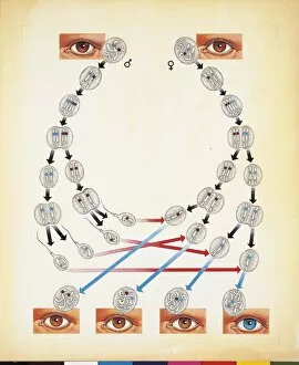
Background imageChromosome Collection: Biological illustration of Human chromosomes combinations with eye-color gene, drawing

Biological illustration of Human chromosomes combinations with eye-color gene, drawing
Biology - Molecular biology - Genetics - Human chromosomes combinations, eye-color gene. Drawing

Background imageChromosome Collection: DNA including double helix, chromosome, cell

DNA including double helix, chromosome, cell

Background imageChromosome Collection: Human karyotype, artwork F007 / 6432

Human karyotype, artwork F007 / 6432
Human karyotype, computer artwork. The karyotype is the complete set of chromosomes in an organism

Background imageChromosome Collection: Human karyotype, artwork F007 / 6431

Human karyotype, artwork F007 / 6431
Human karyotype, computer artwork. The karyotype is the complete set of chromosomes in an organism

Background imageChromosome Collection: Giant chromosomes, SEM

Giant chromosomes, SEM
Giant chromosomes. Colured scanning electron micrograph (SEM) of giant (polytene) chromosomes from a fruit fly (Drosophila busckii)

Background imageChromosome Collection: Chromosomes, artwork F007 / 8518

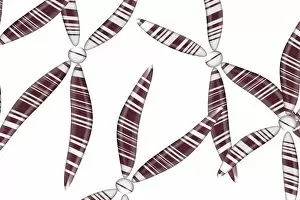
Chromosomes, artwork F007 / 8518
Chromosomes, computer artwork

Background imageChromosome Collection: Chromosomes, artwork F007 / 8517

Chromosomes, artwork F007 / 8517
Chromosomes, computer artwork

Background imageChromosome Collection: Genetic engineering, conceptual image F007 / 8516

Genetic engineering, conceptual image F007 / 8516
Genetic engineering, conceptual composite image

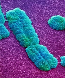
Background imageChromosome Collection: Human chromosome

Human chromosome. Coloured scanning electron micrograph (SEM) of a human chromosome. Chromosomes occur in the nucleus of every cell in the body


All Professionally Made to Order for Quick Shipping

"Unlocking the Mysteries of Life: Exploring the World of Chromosomes" Witness the intricate dance of life as cells divide through mitosis, captured in a stunning light micrograph. Dive into the mesmerizing realm of cell division with a fluorescent micrograph, revealing the vibrant beauty hidden within. Behold a full set of male chromosomes under an SEM, offering a glimpse into our genetic blueprint and diversity. Marvel at the captivating sight of dividing cells, where new life emerges and growth takes place before your eyes. Delve deeper into cell division's enchantment with another fluorescent micrograph that showcases its breathtaking complexity. Unravel the secrets held within chromosome structure through an illuminating illustration that unveils their unique composition. Embark on a journey through DNA replication as bases attach to strands, forming two identical double DNA strands in this biomedical illustration. Explore protein synthesis and ribosome function in cross-section biomedical artwork that reveals how they contribute to cellular processes. Discover the essence of life encapsulated in each chromosome—a testament to our individuality and shared humanity. Traverse across nerve cells like astrocytes while contemplating their integral role in transmitting information throughout our complex nervous system. Immerse yourself in these captivating images—such as human chromosomes observed under an SEM—and let them ignite your curiosity about the wonders concealed within every living organism's DNA structure.