Fraud Blocker Skip to main content

Chromosomes Collection

Chromosomes are the intricate threads that hold the blueprint of life within our cells

Background imageChromosomes Collection: X and Y chromosomes

X and Y chromosomes, coloured scanning electron micrograph (SEM). Humans have 46 chromosomes in total: 23 inherited from the mother and 23 from the father

Background imageChromosomes Collection: Mitosis, light micrograph

Mitosis, light micrograph
Mitosis. Confocal light micrograph of the stages of mitosis (nuclear division) and cytokinesis (cell division). During mitosis the nuclear envelope disintegrates (3rd image) and the chromosomes (blue)

Background imageChromosomes Collection: Cell division, fluorescent micrograph

Cell division, fluorescent micrograph
Cell division. Immunofluorescent light micrograph of a human epithelial cell (centre) during the late anaphase stage of mitosis

Background imageChromosomes Collection: Biomedical illustration of DNA Replication as bases attach to strand with two newly formed strands

Biomedical illustration of DNA Replication as bases attach to strand with two newly formed strands twist eventually producing two new identical double DNA strands

Background imageChromosomes Collection: Full set of male chromosomes, SEM

Full set of male chromosomes, SEM
Set of human chromosomes, coloured scanning election micrograph (SEM). Chromosomes are a packaged form of the genetic material deoxyribonucleic acid (DNA)

Background imageChromosomes Collection: Chromosome

Chromosome. Computer artwork of a condensed chromosome. Chromosomes, which consist of two identical chromatids joined at a centromere (centre)

Background imageChromosomes Collection: Dividing cells

Dividing cells. Computer artwork of a spiral of cells during mitosis (nuclear division). During mitosis two daughter nuclei are formed from one parent nucleus

Background imageChromosomes Collection: Cell division, fluorescent micrograph

Cell division, fluorescent micrograph
Cell division. Immunofluorescent light micrograph of a human epithelial cell (centre) during the interphase stage of mitosis

Background imageChromosomes Collection: Cross section biomedical illustration of protein synthesis and ribosome

Cross section biomedical illustration of protein synthesis and ribosome

Background imageChromosomes Collection: Astrocyte nerve cell

Astrocyte nerve cell. Fluorescent light micrograph of an astrocyte cell from a human brain. Intermediate filaments (IFs), part of the cells cytoskeleton, have been dyed green

Background imageChromosomes Collection: X and Y chromosomes

X and Y chromosomes, computer artwork. Humans have 46 chromosomes in total: 23 inherited from the mother and 23 from the father. These sex chromosomes determine the individuals gender

Background imageChromosomes Collection: Human chromosomes, SEM C013 / 5002

Human chromosomes, SEM C013 / 5002
Human chromosomes. Coloured scanning electron micrograph (SEM) of human chromosomes prepared with the harlequin staining technique

Background imageChromosomes Collection: Plant cell mitosis, light micrograph

Plant cell mitosis, light micrograph
Plant cell mitosis. Light micrograph of root tip cells from an onion (Allium sp.) undergoing cell division (mitosis). From top left to bottom right

Background imageChromosomes Collection: Male sex chromosomes, SEM

Male sex chromosomes, SEM
Male sex chromosomes. Coloured scanning electron micrograph (SEM) of human X (centre) and Y (upper left) sex chromosomes. Each chromosome has replic- ated to form two identical strands (chromatids)

Background imageChromosomes Collection: Biomedical illustration of DNA Replication

Biomedical illustration of DNA Replication

Background imageChromosomes Collection: Biomedical illustration of protein synthesis within DNA

Biomedical illustration of protein synthesis within DNA

Background imageChromosomes Collection: Double Helix of Human DNA

Double Helix of Human DNA

Background imageChromosomes Collection: Artwork of DNA structure

Artwork of DNA structure

Background imageChromosomes Collection: Giant chromosomes, SEM

Giant chromosomes, SEM
Giant chromosomes. Colured scanning electron micrograph (SEM) of giant (polytene) chromosomes from a fruit fly (Drosophila busckii)

Background imageChromosomes Collection: Human chromosome

Human chromosome. Coloured scanning electron micrograph (SEM) of a human chromosome. Chromosomes occur in the nucleus of every cell in the body

Background imageChromosomes Collection: Giant chromosomes, SEM P657 / 0034

Giant chromosomes, SEM P657 / 0034
Giant chromosomes. Coloured scanning electron micrograph (SEM) of giant (polytene) chromosomes from a fruit fly (Drosophila busckii)

Background imageChromosomes Collection: LM of Drosophila giant (polytene) chromosome

LM of Drosophila giant (polytene) chromosome
Giant chromosome. Coloured computer-enhanced light micrograph of a giant (polytene) chromosome in a salivary gland cell of the fruit fly Drosophila funebris

Background imageChromosomes Collection: LM of giant polytene chromosomes, Drosophila

LM of giant polytene chromosomes, Drosophila
Light micrograph of stained giant polytene chromosomes of Drosophila funebris. These chromosomes are found in the salivary glands of D.funebris and many other fly species

Background imageChromosomes Collection: Targeted gene on a chromosome, SEM

Targeted gene on a chromosome, SEM
Targeted gene on a human chromosome. Composite coloured scanning electron micrograph (SEM) of a targeted gene (yellow) on one arm of a human chromosome

Background imageChromosomes Collection: Chromosomes, SEM

Chromosomes, SEM
Chromosomes. Coloured scanning electron micrograph (SEM) of two chromosomes. The SEM is overlaid on a DNA autoradiogram background

Background imageChromosomes Collection: Giant chromosomes, light micrograph P657 / 0038

Giant chromosomes, light micrograph P657 / 0038
Giant chromosomes. Light micrograph of giant (polytene) chromosomes from a fruit fly (Drosophila busckii). These chromosomes are formed from repeated rounds of DNA (deoxyribonucleic acid)

Background imageChromosomes Collection: Giant chromosomes, light micrograph P657 / 0037

Giant chromosomes, light micrograph P657 / 0037
Giant chromosomes. Light micrograph of giant (polytene) chromosomes from a fruit fly (Drosophila busckii). These chromosomes are formed from repeated rounds of DNA (deoxyribonucleic acid)

Background imageChromosomes Collection: E. coli Holliday junction complex C014 / 0878

E. coli Holliday junction complex C014 / 0878
E. coli Holliday junction complex. Molecular model of a RuvA protein (dark pink) in complex with a Holliday junction between homologous strands of DNA (deoxyribonucleic acid)

Background imageChromosomes Collection: Gene expression, artwork

Gene expression, artwork
Gene expression. Computer artwork showing the process of transcription, the first stage or gene expression. Here, a chromosome (distance)

Background imageChromosomes Collection: DNA Holliday junction complex F006 / 9334

DNA Holliday junction complex F006 / 9334
DNA Holliday junction complex. Molecular model of the enzyme FLP recombinase in complex with a Holliday junction between homologous strands of DNA (deoxyribonucleic acid)

Background imageChromosomes Collection: E coli Holliday junction complex F006 / 9261

E coli Holliday junction complex F006 / 9261
E. coli Holliday junction complex. Molecular model of a RuvA protein (red) in complex with a Holliday junction between homologous strands of DNA (deoxyribonucleic acid, blue) from an E

Background imageChromosomes Collection: Roundworm germ cells, light micrograph C016 / 9538

Roundworm germ cells, light micrograph C016 / 9538
Roundworm germ cells. Light micrograph of germ cells from a roundworm (Ascaris sp.), undergoing mitosis (nuclear division)

Background imageChromosomes Collection: Genetic fingerprints, conceptual artwork C016 / 7521

Genetic fingerprints, conceptual artwork C016 / 7521
Genetic fingerprints, conceptual computer artwork

Background imageChromosomes Collection: Chromosome segregation protein

Chromosome segregation protein, molecular model. This proteins function is to aid the process of chromosome segregation during cell division and replication

Background imageChromosomes Collection: Male XY chromosomes, artwork

Male XY chromosomes, artwork. The Y chromosome (right) is found in males and is shorter than the X chromosome (upper left)

Background imageChromosomes Collection: Dividing cell, TEM

Dividing cell, TEM
Dividing cell. Transmission electron micrograph (TEM) of a section through a cell dividing through the process of mitosis

Background imageChromosomes Collection: Drug effect on viruses, conceptual image C016 / 6253

Drug effect on viruses, conceptual image C016 / 6253
Drug effect on viruses, conceptual image. Computer artwork showing a single strand of DNA (deoxyribonucleic acid, spiral, centre), red blood cells (pink), virus particles (virions, green, small)

Background imageChromosomes Collection: Chromosomes, light micrograph C016 / 6354

Chromosomes, light micrograph C016 / 6354
Chromosomes, light micrograph. This micrograph was obtained in 1934 during studies of chromosome structure in the salivary glands of Sciara (a genus of fungus gnats)

Background imageChromosomes Collection: Meiosis, illustration C018 / 0803

Meiosis, illustration C018 / 0803
Meiosis. Illustration of a cell during metaphase (I) of meiosis (gamete formation). During meiosis four daughter nuclei are formed from one parent nucleus after two stages of nuclear division

Background imageChromosomes Collection: Gene expression, conceptual illustration C018 / 0746

Gene expression, conceptual illustration C018 / 0746
Gene expression, conceptual illustration. Every cell in an organism contains every single gene that makes up the organisms genome. However, they are not all active (expressed) in each cell

Background imageChromosomes Collection: Genetic karyotype analysis C019 / 0270

Genetic karyotype analysis C019 / 0270
MODEL RELEASED. Genetic karyotype analysis. Analyst in a genetics laboratory studying the results after DNA (deoxyribonucleic acid) was isolated from a sample

Background imageChromosomes Collection: Genetic karyotype analysis C019 / 0271

Genetic karyotype analysis C019 / 0271
Genetic karyotype analysis. Analyst in a genetics laboratory studying the results after DNA (deoxyribonucleic acid) was isolated from a sample

Background imageChromosomes Collection: Genetic karyotype analysis C019 / 0268

Genetic karyotype analysis C019 / 0268
MODEL RELEASED. Genetic karyotype analysis. Analyst in a genetics laboratory studying the results after DNA (deoxyribonucleic acid) was isolated from a sample

Background imageChromosomes Collection: Genetic karyotype analysis C019 / 0269

Genetic karyotype analysis C019 / 0269
MODEL RELEASED. Genetic karyotype analysis. Analyst in a genetics laboratory studying the results after DNA (deoxyribonucleic acid) was isolated from a sample

Background imageChromosomes Collection: E. coli Holliday junction complex

E. coli Holliday junction complex. Molecular model of a RuvA protein (red) in complex with a Holliday junction between homologous strands of DNA (deoxyribonucleic acid, brown and orange) from an E

Background imageChromosomes Collection: DNA quadruplex, molecular model

DNA quadruplex, molecular model. This dimeric quadruplex of DNA (deoxyribonucleic acid) is thought to form as part of telomeres

Background imageChromosomes Collection: DNA Holliday junction, molecular model C014 / 3090

DNA Holliday junction, molecular model C014 / 3090
DNA Holliday junction. Molecular model of a Holliday junction (centre) between homologous strands of DNA (deoxyribonucleic acid)

Background imageChromosomes Collection: Chromosomes, artwork C013 / 9968

Chromosomes, artwork C013 / 9968
Chromosomes. Computer artwork of human chromosomes. Chromosomes are composed of deoxyribonucleic acid (DNA) that contain sections, called genes, which encode the bodys genetic information


All Professionally Made to Order for Quick Shipping

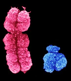
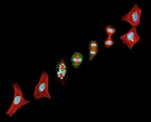
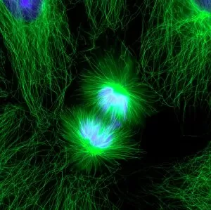
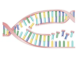
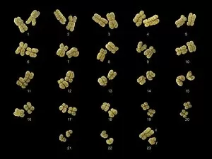
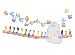
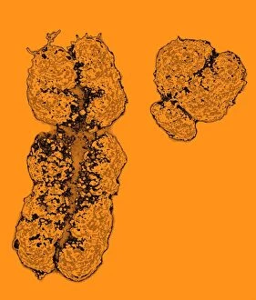
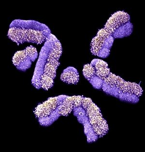
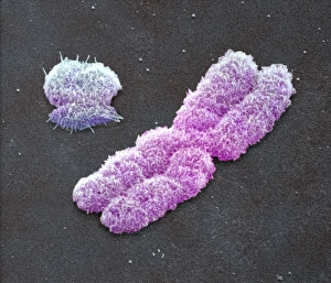
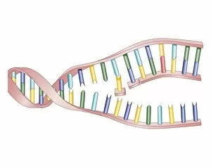
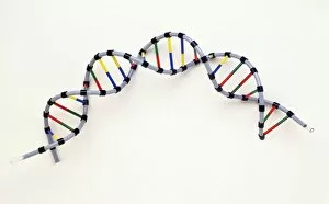
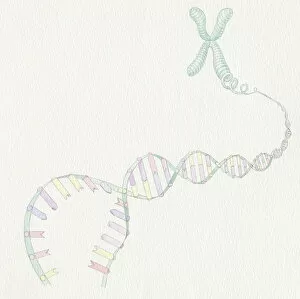
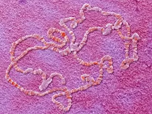
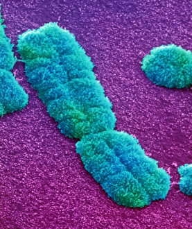
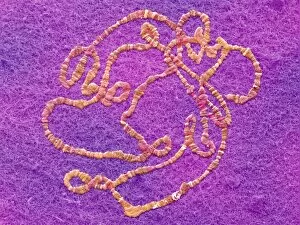
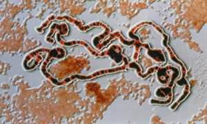
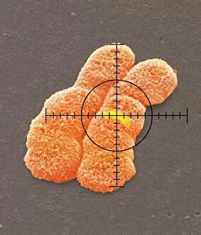
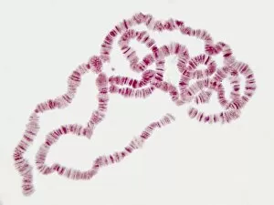
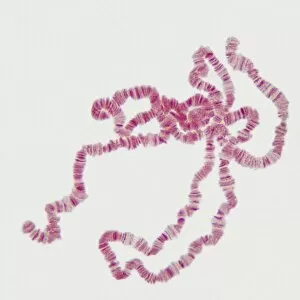
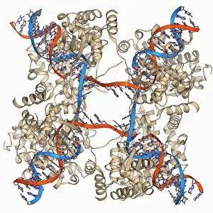
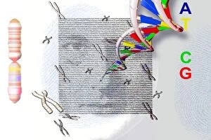
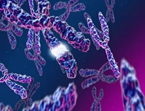
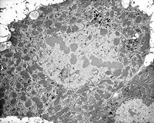
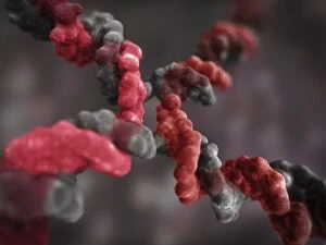
Chromosomes are the intricate threads that hold the blueprint of life within our cells. Among them, the X and Y chromosomes determine our biological sex, with males having one X and one Y chromosome. Through processes like mitosis, these chromosomes replicate themselves to ensure proper cell division. Under a light micrograph, we can witness this incredible phenomenon in action. In fluorescent micrographs capturing cell division, vibrant colors illuminate the intricate dance as they separate into two new cells. This mesmerizing display showcases the beauty hidden within our bodies. Examining a full set of male chromosomes under a scanning electron microscope (SEM) reveals their unique structure and arrangement. These microscopic images provide us with invaluable insights into our genetic makeup. Dividing cells captured in fluorescent micrographs remind us of the constant renewal happening inside us. Each split brings forth new possibilities for growth and development. Biomedical illustrations vividly depict DNA replication—a crucial process where bases attach to strands forming two identical double DNA strands. The twisting motion symbolizes how life's building blocks come together to create something extraordinary. A cross-sectional biomedical illustration unveils protein synthesis and ribosome activity—essential components for cellular function. This complex mechanism ensures that proteins are produced accurately according to instructions encoded in our chromosomes. The image of a single chromosome evokes wonder at its intricacy—an elegant structure carrying vital information that shapes who we are at a fundamental level. Astrocyte nerve cells intertwine with our understanding of chromosomes' role beyond reproduction—they play critical roles in brain function and communication between neurons. Under SEM imaging techniques, human chromosomes reveal their awe-inspiring complexity up close—each strand holding countless secrets waiting to be unraveled by scientists seeking answers about life itself.