
Centromere Collection
The centromere, a crucial component of chromosomes, plays a vital role in cell division and genetic inheritance


All Professionally Made to Order for Quick Shipping
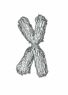
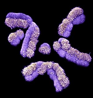
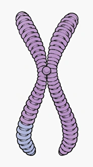
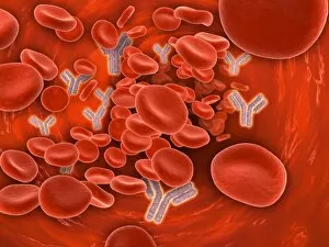
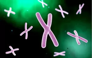
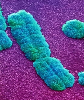
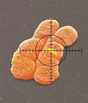
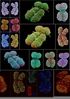
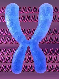
The centromere, a crucial component of chromosomes, plays a vital role in cell division and genetic inheritance. Found at the center of each chromosome, it serves as an anchor point for spindle fibers during mitosis and meiosis. This microscopic view showcases the intricate structure of human chromosomes, with their distinct chromatids and prominent centromeres. As we zoom in further, we discover the fascinating telomeres highlighted at the tips of these chromosomes – protective caps that prevent DNA damage and ensure stability. The conceptual images depict various aspects related to chromosomes: from their formation inside astrocyte nerve cells to their presence within our bloodstream. These captivating visuals remind us of the complexity and beauty hidden within our genetic makeup. Understanding the centromere's function is essential for comprehending how DNA is faithfully replicated and passed on from one generation to another – a remarkable process that shapes who we are as individuals.