Mounted Print > Popular Themes > Human Body
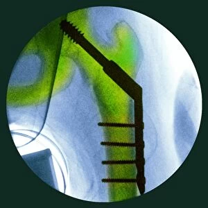
Mounted Print : Total hip replacement, X-ray


![]()
Mounted Prints from Science Photo Library
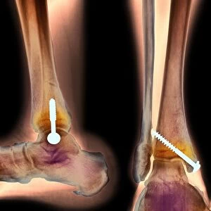
Total hip replacement, X-ray
Total hip replacement. Coloured frontal X-ray of a female pelvis with a total hip replacement (white, lower right). The replacement hip consists of a ball and shaft, embedded in the femur (thigh bone, running up from bottom right), which moves freely in the prosthetic socket implanted in the pelvis
Science Photo Library features Science and Medical images including photos and illustrations
Media ID 6391577
© MIRIAM MASLO/SCIENCE PHOTO LIBRARY
Artificial Implant Ball And Socket Bones Degeneration Degenerative Devices False Colour Femur Frontal Implants Joint Osteology Procedure Prosthesis Prosthetic Device Radiography Replaced Shaft Surgery Surgical Thigh Thigh Bone Total Hip Replacement Treatment X Ray X Ray Machine Abnormal Condition Disorder False Coloured Health Care Pelvis Right Leg Unhealthy
10"x8" Mount with 8"x6" Print
Discover the transformative power of our Media Storehouse Mounted Photos with this captivating image of a total hip replacement X-ray. Witness the intricacy and innovation of medical advancements brought to life in vibrant color. This coloured frontal X-ray of a female pelvis reveals a total hip replacement, where a white ball and shaft replace the natural hip joint. A must-have addition to any medical or educational space, this mounted photo is sure to inspire and inform.
Printed on 8"x6" paper and suitable for use in a 10"x8" frame (frame not included). Prints are mounted with card both front and back. Featuring a custom cut aperture to match chosen image. Professional 234gsm Fujifilm Crystal Archive DP II paper.
Photo prints supplied in custom cut card mount ready for framing
Estimated Image Size (if not cropped) is 18.3cm x 15.2cm (7.2" x 6")
Estimated Product Size is 25.4cm x 20.3cm (10" x 8")
These are individually made so all sizes are approximate
Artwork printed orientated as per the preview above, with landscape (horizontal) orientation to match the source image.
EDITORS COMMENTS
This print showcases a total hip replacement procedure, providing a detailed and colorful frontal view of a female pelvis. The image highlights the innovative technology used in modern medicine to address joint disorders and improve overall health. In this case, a white prosthetic socket is implanted in the pelvis, serving as an artificial implant for the replaced hip joint. The complexity of this surgical intervention becomes apparent as we observe the ball and shaft component embedded within the femur, which allows for smooth movement and functionality. This cutting-edge medical device enables individuals suffering from degenerative conditions or other hip-related disorders to regain their mobility and enhance their quality of life. The vibrant green hues applied to this X-ray image add depth and clarity, aiding healthcare professionals in accurately diagnosing any abnormalities or degeneration present within the bones. With advancements in radiography techniques and equipment like x-ray machines, doctors can now provide precise treatment plans tailored to each patient's unique needs. As we delve into this remarkable photograph by Science Photo Library, it serves as a powerful reminder of how science intersects with human anatomy to create groundbreaking solutions that positively impact countless lives worldwide.
MADE IN THE UK
Safe Shipping with 30 Day Money Back Guarantee
FREE PERSONALISATION*
We are proud to offer a range of customisation features including Personalised Captions, Color Filters and Picture Zoom Tools
SECURE PAYMENTS
We happily accept a wide range of payment options so you can pay for the things you need in the way that is most convenient for you
* Options may vary by product and licensing agreement. Zoomed Pictures can be adjusted in the Basket.