Fraud Blocker Skip to main content

Pelvis Collection

The pelvis: A vital foundation of the human body's intricate design

Background imagePelvis Collection: Human Anatomy - Backbone including Ribs and Pelvis

Human Anatomy - Backbone including Ribs and Pelvis
Vintage engraving of the human Backbone including Ribs and Pelvis. 19th Century

Background imagePelvis Collection: Hip replacement, artwork

Hip replacement, artwork
Hip replacement. Artwork of a total hip replacement. The prosthetic hip joint consists of a metal shaft and ball (pink) attached to the top of the femur (thigh bone) and a plastic socket (white)

Background imagePelvis Collection: Cardiovascular system, historical artwork

Cardiovascular system, historical artwork. 19th Century hand coloured lithographic print showing the blood vessels (red and blue) of the human neck and torso

Background imagePelvis Collection: Male and Female pelvis engraving 1896

Male and Female pelvis engraving 1896
Atlas d anatomie descriptive du corps humain C. Bonamy - Paul Broca Victor Masson et Fils Paris 1866

Background imagePelvis Collection: Total hip replacement, X-ray

Total hip replacement, X-ray
Total hip replacement. Coloured frontal X-ray of a female pelvis with a total hip replacement (white, lower right). The replacement hip consists of a ball and shaft

Background imagePelvis Collection: Upper body skeleton, computer artwork

Upper body skeleton, computer artwork
Upper body skeleton. Computer artwork of a human skeleton showing the bones of the upper body. At the top is the base of the skull and the jaw bone

Background imagePelvis Collection: Diagram of the bones of the right leg and hip

Diagram of the bones of the right leg and hip
Diagram of the bones of the right leg, showing the joint with the pelvis at the hip

Background imagePelvis Collection: Normal spine, X-ray

Normal spine, X-ray
Normal spine. Coloured profile X-ray showing bones of the spine (vertebrae, centre). The front of the body is at right. The ribs (upper right) and the pelvis (bottom) are also seen

Background imagePelvis Collection: Hip replacement, artwork

Hip replacement, artwork
Hip replacement. Artwork of a prosthetic hip joint socket in a pelvis. The prosthetic femur (thigh bone) head will fit into this socket, forming the hip joint

Background imagePelvis Collection: Cross section biomedical illustration of iliac crest site on pelvis

Cross section biomedical illustration of iliac crest site on pelvis

Background imagePelvis Collection: The human body with superimposed colored plates by Julien Bougle

The human body with superimposed colored plates by Julien Bougle
The human body with superimposed colored plates, by Julien Bougle, circa 1899

Background imagePelvis Collection: Human arm musculature, artwork F007 / 1810

Human arm musculature, artwork F007 / 1810
Human arm musculature, computer artwork

Background imagePelvis Collection: Osteoarthritis of the hip, X-ray

Osteoarthritis of the hip, X-ray
Coloured X-ray (front view) of the pelvis in a 57 year old patient showing osteoarthritis of the hip joint (at left). The bone in the head of the femur (thigh bone)

Background imagePelvis Collection: Slipped disc

Slipped disc. Spinal model demonstrating a slipped intervertebral disc. The slipped disc is the small bulge (red, centre), near the base of the spine

Background imagePelvis Collection: Slipped intervertebral disc, artwork

Slipped intervertebral disc, artwork
Slipped intervertebral disc, computer artwork. Intervertebral discs (red) are pads of cartilage between the bones (vertebrae) of the spine

Background imagePelvis Collection: Front view of the female anatomy hilighting the endocrine system

Front view of the female anatomy hilighting the endocrine system
Brain, Anatomy, Diagram, Heart, Illustration, Bone, Shoulder, Medical, Uterus, Biology, Pelvis, Femur, See Through, Vertical, Artwork, Female Likeness, Color Image, Front View

Background imagePelvis Collection: Digital illustration of dislocated hip

Digital illustration of dislocated hip
Anatomy, Biomedical Illustration, Close-Up, Cross Section, Digitally Generated, Dislocation, Healthcare and Medicine, Hip Bone, Human Bone, Illustration and Painting, Illustrative Technique

Background imagePelvis Collection: Anterior view of pelvis with hip bones showing arthritis and osteophytes on femoral heads

Anterior view of pelvis with hip bones showing arthritis and osteophytes on femoral heads
Horizontal, Anatomy, Diagram, Illustration, Bone, Disease, Medical, Hip, Biology, Pelvis, Femur, Cut Out, Arthritis, White Background, Artwork, Color Image, Front View, Healthcare And Medicine

Background imagePelvis Collection: Normal anterior view of pelvis with hip bones

Normal anterior view of pelvis with hip bones
Horizontal, Anatomy, Diagram, Illustration, Bone, Medical, Hip, Biology, Pelvis, Femur, Cut Out, White Background, Artwork, Color Image, Front View, Healthcare And Medicine, The Human Body

Background imagePelvis Collection: Front view of a woman nine months pregnant (baby phantomed within) ready for delivery

Front view of a woman nine months pregnant (baby phantomed within) ready for delivery
Detail, Baby, Anatomy, Diagram, Mother, Illustration, Bone, Birth, Medical, Fetus, Pregnant, Pregnancy, Biology, Pelvis, Femur, Development, Abdomen, Anterior, See Through, Cut Out, White Background

Background imagePelvis Collection: Repetitive activities like aerobics that involve lifting up the knee can cause trauma to

Repetitive activities like aerobics that involve lifting up the knee can cause trauma to
Detail, Anatomy, Diagram, Muscle, Pain, Illustration, Bone, Trauma, Injury, Medical, Biology, Pelvis, Femur, See Through, Cut Out, White Background, Artwork, Color Image, Front View, Close Up

Background imagePelvis Collection: Snapping hip syndrome occurs when the iliopsoas tendon subluxes over the greater

Snapping hip syndrome occurs when the iliopsoas tendon subluxes over the greater
Horizontal, Anatomy, Diagram, Muscle, Illustration, Bone, Medical, Hip, Biology, Pelvis, Femur, See Through, Rear View, Cut Out, Arthritis, White Background, Artwork, Color Image, Front View

Background imagePelvis Collection: Normal abdomen, X-ray

Normal abdomen, X-ray
Normal abdomen. X-ray of the abdomen of a 20 year old female

Background imagePelvis Collection: Study of the Human Figure, Anterior View, from A Comparative Anatomical Exposition of

Study of the Human Figure, Anterior View, from A Comparative Anatomical Exposition of the Structure of the Human Body
XYC257409 Study of the Human Figure, Anterior View, from A Comparative Anatomical Exposition of the Structure of the Human Body with that of a Tiger and a Common Fowl

Background imagePelvis Collection: Normal anatomy of an open hip showing the articular surface of the femur

Normal anatomy of an open hip showing the articular surface of the femur
Detail, Anatomy, Diagram, Illustration, Bone, Surface, Medical, Hip, Biology, Pelvis, Femur, Cut Out, White Background, Vertical, Artwork, Color Image, Close Up, Healthcare And Medicine

Background imagePelvis Collection: Normal anterior view of a pelvis

Normal anterior view of a pelvis
Horizontal, Anatomy, Diagram, Illustration, Bone, Medical, Hip, Biology, Pelvis, Cut Out, White Background, Artwork, Color Image, Front View, Healthcare And Medicine, The Human Body, No People

Background imagePelvis Collection: Anterior view of human skeletal system, with labels

Anterior view of human skeletal system, with labels

Background imagePelvis Collection: Anatomy of male human skeleton, side view and perspective view

Anatomy of male human skeleton, side view and perspective view

Background imagePelvis Collection: The Elvis

The Elvis
Hanna Lee Tidd

Background imagePelvis Collection: The Elvis

The Elvis
Hanna Lee Tidd

Background imagePelvis Collection: Ligaments of the human body (engraving)

Ligaments of the human body (engraving)
5216894 Ligaments of the human body (engraving) by German School, (19th century); Private Collection; (add.info.: Ligaments of the human body)

Background imagePelvis Collection: Diseases of the Stomach and Kidneys (colour litho)

Diseases of the Stomach and Kidneys (colour litho)
6006706 Diseases of the Stomach and Kidneys (colour litho) by European School, (19th century); Private Collection; (add.info.: Diseases of the Stomach and Kidneys)

Background imagePelvis Collection: Skeleton of an Eagle, after Milne-Edwards (engraving)

Skeleton of an Eagle, after Milne-Edwards (engraving)
966625 Skeleton of an Eagle, after Milne-Edwards (engraving) by English School, (19th century); Private Collection; (add.info.: Skeleton of an Eagle, after Milne-Edwards)

Background imagePelvis Collection: Man on horseback, exhibiting side view of pelvis (engraving)

Man on horseback, exhibiting side view of pelvis (engraving)
6001158 Man on horseback, exhibiting side view of pelvis (engraving) by English School, (19th century); Private Collection; (add.info.: Man on horseback, exhibiting side view of pelvis)

Background imagePelvis Collection: Section of human kidney (litho)

Section of human kidney (litho)
6013564 Section of human kidney (litho) by English School, (20th century); Private Collection; (add.info.: Section of human kidney)

Background imagePelvis Collection: Anatomical Study of the Human Skeleton, 1896 (chromolithograph)

Anatomical Study of the Human Skeleton, 1896 (chromolithograph)
2622453 Anatomical Study of the Human Skeleton, 1896 (chromolithograph) by American School, (19th century); Private Collection; (add.info.: Antique anatomical study of the human skeleton)

Background imagePelvis Collection: Temporary Lateral Curvature of the Spine in a Normal Child, Child Sitting Erect

Temporary Lateral Curvature of the Spine in a Normal Child, Child Sitting Erect
3633313 Temporary Lateral Curvature of the Spine in a Normal Child, Child Sitting Erect, Lateral Curvature of the Spine (b/w photo) by English Photographer

Background imagePelvis Collection: Side view of pelvis, showing Levator ani (engraving)

Side view of pelvis, showing Levator ani (engraving)
3634198 Side view of pelvis, showing Levator ani (engraving) by English School, (19th century); Private Collection; (add.info.: Side view of pelvis, showing Levator ani)

Background imagePelvis Collection: Midwifery, illustration from Encyclopedia Britannica, published in Edinburgh

Midwifery, illustration from Encyclopedia Britannica, published in Edinburgh
DGC398035 Midwifery, illustration from Encyclopedia Britannica, published in Edinburgh, 1798 (copperplate engraving) by Bell, Alexander (fl)

Background imagePelvis Collection: Two studies of a hanging skeleton (pen & ink with wash)

Two studies of a hanging skeleton (pen & ink with wash)
AII80166 Two studies of a hanging skeleton (pen & ink with wash) by Vinci, Leonardo da (1452-1519); Gabinetto dei Disegni e Stampe, Galleria Degli Uffizi, Florence, Tuscany, Italy; Italian

Background imagePelvis Collection: Front view of a body showing a total hip replacement

Front view of a body showing a total hip replacement
Detail, Anatomy, Diagram, Illustration, Bone, Disease, Medical, Hip, Biology, Pelvis, Femur, Acetabulum, Surgery, See Through, Cut Out, White Background, Vertical, Artwork, Color Image, Front View

Background imagePelvis Collection: Normal posterior view of the hip bones and gluteus maximus muscle including the lumbar

Normal posterior view of the hip bones and gluteus maximus muscle including the lumbar
Detail, Anatomy, Diagram, Muscle, Illustration, Bone, Medical, Hip, Biology, Pelvis, Femur, See Through, Rear View, Cut Out, White Background, Artwork, Color Image, Close Up, Healthcare And Medicine

Background imagePelvis Collection: Normal posterior view of the back highlighting the multifidus muscle

Normal posterior view of the back highlighting the multifidus muscle
Detail, Anatomy, Diagram, Muscle, Illustration, Ribs, Bone, Medical, Biology, Pelvis, See Through, Rear View, Cut Out, White Background, Artwork, Color Image, Close Up, Healthcare And Medicine

Background imagePelvis Collection: Human rib cage, jaw bones, neck vertabrae leading to sternum, collar bones and ribcage

Human rib cage, jaw bones, neck vertabrae leading to sternum, collar bones and ribcage, spinal column continuing to pelvis, front view

Background imagePelvis Collection: Anatomy skeleton

Anatomy skeleton
Diagram of the human skeleton from front and back

Background imagePelvis Collection: Female body with bone skeleton and internal organs superimposed

Female body with bone skeleton and internal organs superimposed
Anatomy of female body with bone skeleton and all internal organs superimposed

Background imagePelvis Collection: Anatomy of female muscular system, back view

Anatomy of female muscular system, back view

Background imagePelvis Collection: Anterior view of human pelvis, with labels

Anterior view of human pelvis, with labels


All Professionally Made to Order for Quick Shipping

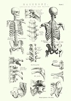
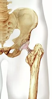
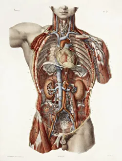
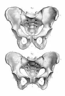
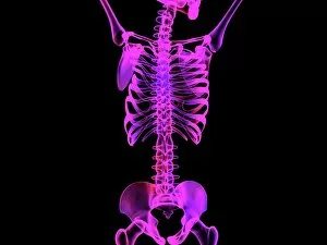
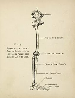
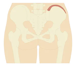
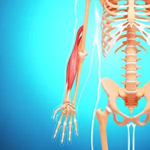
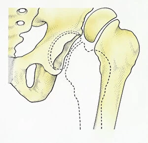
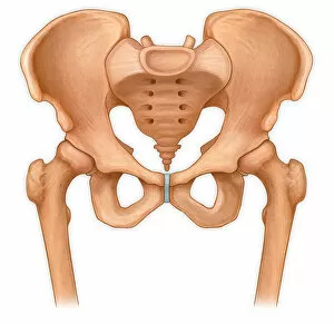
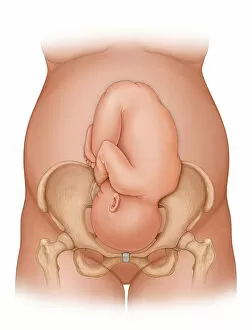
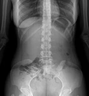
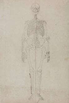
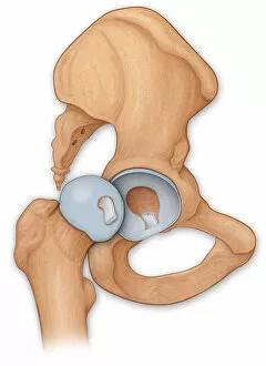
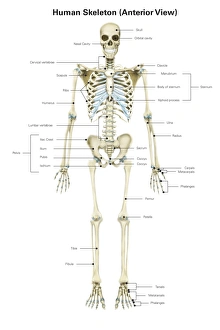
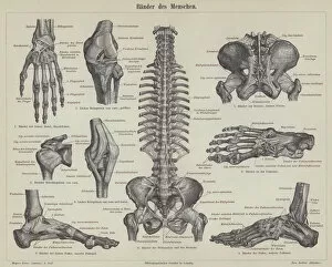
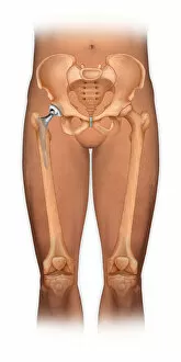
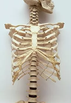
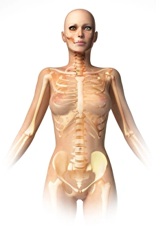
The pelvis: A vital foundation of the human body's intricate design. From its role in supporting the backbone, including ribs and pelvis, to its distinct differences between male and female anatomy depicted in a captivating 1896 engraving. Explore the connection between the cardiovascular system and this historical artwork, showcasing the significance of every bone and joint. Witness the transformative power of hip replacement through stunning artistic representations and X-rays capturing both total hip replacements and normal spines. Delve into computer artwork depicting an upper body skeleton, highlighting how each element contributes to our overall structure. Study a detailed diagram showcasing the bones of the right leg and hip, unraveling their complexity with precision. Marvel at Julien Bougle's masterpiece - a human body adorned with superimposed colored plates that reveal hidden wonders within us all. Discover how osteoarthritis affects this crucial region through an enlightening X-ray image. Finally, admire F007/1810's exquisite artwork unveiling the intricacies of human arm musculature – a testament to our remarkable physical capabilities.