
Degenerative Collection
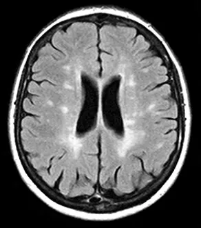
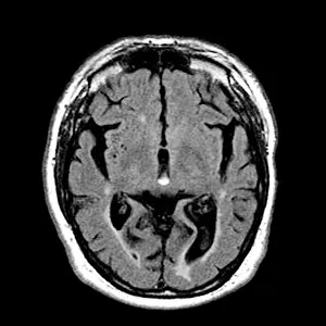
"Degenerative conditions captured through medical imaging: Total hip replacement, X-ray; Multiple sclerosis, SEM; Alcoholic dementia, MRI scan


All Professionally Made to Order for Quick Shipping
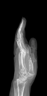
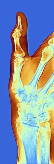
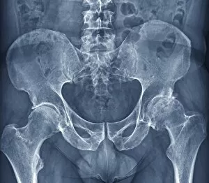
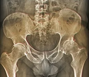
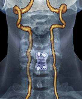
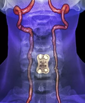
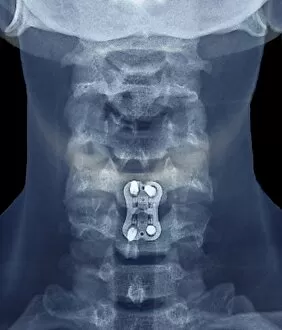
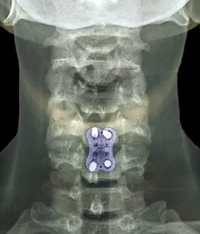
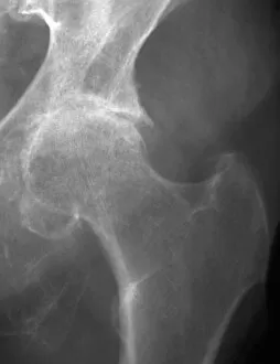
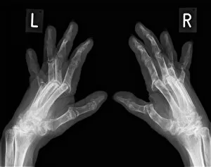
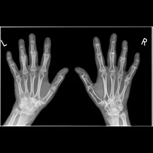
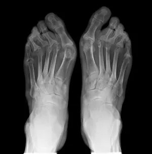
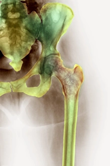
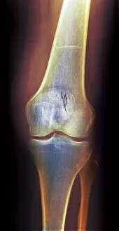
"Degenerative conditions captured through medical imaging: Total hip replacement, X-ray; Multiple sclerosis, SEM; Alcoholic dementia, MRI scan. These images reveal the effects of degeneration on various parts of the body. Arthritis of the neck and arthrosis of the hand are evident in X-rays C017 / 7389, F006 / 4616, F006 / 4595, and C017 / 7171 respectively. Osteoarthritis takes its toll on the hip as seen in X-rays F006 / 3744 and F006 / 3745. The intricate details of an arthritic hand are also visible in an X-ray image. These visuals provide valuable insights into degenerative conditions that affect individuals' quality of life. "