
X Ray Collection
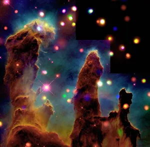
"Unveiling the Hidden World: Exploring the Marvels of X-ray Imaging" Pillars of Creation: Witness the breathtaking beauty of celestial formations through an X-ray lens


All Professionally Made to Order for Quick Shipping
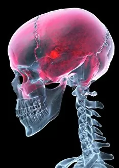
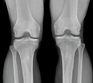
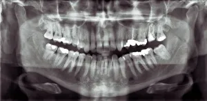
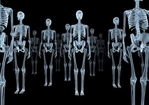
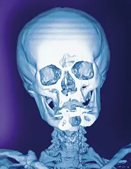
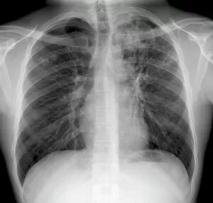
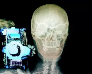
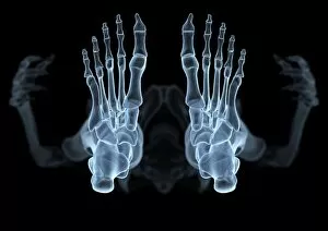
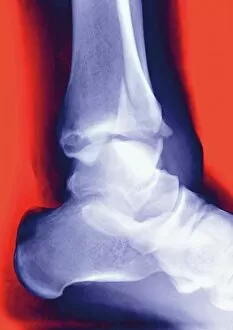
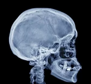
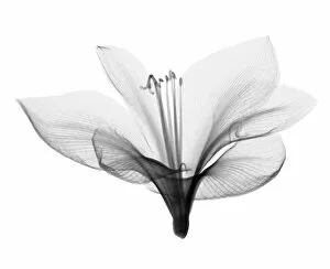
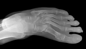
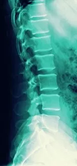
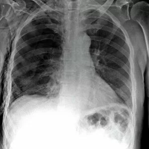
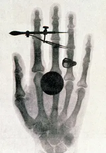
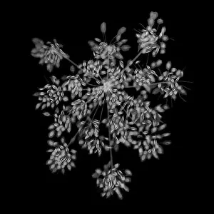
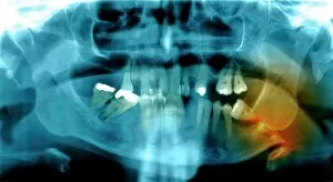
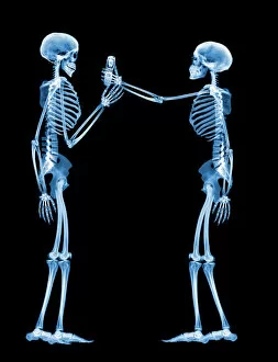
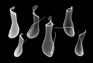
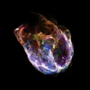
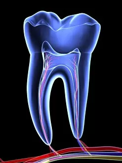
"Unveiling the Hidden World: Exploring the Marvels of X-ray Imaging" Pillars of Creation: Witness the breathtaking beauty of celestial formations through an X-ray lens, revealing a mesmerizing view beyond what meets the eye. Normal Knees, X-rayed: Delve into the intricate structure of our joints as we peer beneath the surface to understand how our knees function in perfect harmony. Panoramic Dental X-ray: Step into a world where dentistry meets technology, capturing a comprehensive image that allows for precise diagnosis and treatment planning. Person with a Camera, X-ray Vision: Unleash your inner superhero as we explore how modern photography techniques merge with X-rays to capture stunning images from within. Tendinitis of the Shoulder, Exposed by X-ray: Discover how this powerful imaging tool helps healthcare professionals identify and diagnose shoulder conditions like tendinitis with accuracy and efficiency. Total Hip Replacement Revealed by X-ray: Journey through medical advancements as we uncover how an artificial hip joint is seamlessly integrated into one's body using cutting-edge surgical techniques guided by detailed x-rays. Normal Skull Underneath It All - An Intriguing Peek Through an X-ray Lens: Explore the hidden secrets concealed within our skulls as we unravel their fascinating anatomy through x-rays. Tuberculosis Unmasked - A Diagnostic Breakthrough via X-rays: Shedding light on one of humanity's oldest diseases, witness how tuberculosis is detected and monitored using specialized x-rays that aid in early detection and effective treatment strategies. Spiral Galaxy M81 - A Cosmic Symphony Captured in Composite Image Using Advanced Radiography Techniques: Embark on an interstellar journey to marvel at this captivating spiral galaxy captured through innovative composite imaging methods involving x-rays and other astronomical data sources. Skeletons Transformed - Artistic Expression Meets Medical Insight in Striking X-ray Artwork.