Mounted Print > Animals > Mammals > Echimyidae > Medius
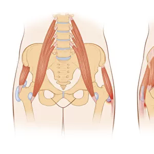
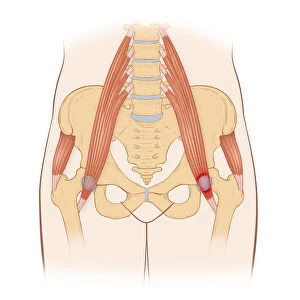
Mounted Print : Snapping hip syndrome occurs when the iliopsoas tendon subluxes over the greater


![]()
Mounted Prints from Fine Art Storehouse
Snapping hip syndrome occurs when the iliopsoas tendon subluxes over the greater
Horizontal, Anatomy, Diagram, Muscle, Illustration, Bone, Medical, Hip, Biology, Pelvis, Femur, See Through, Rear View, Cut Out, Arthritis, White Background, Artwork, Color Image, Front View, Healthcare And Medicine, The Human Body, Human Anatomy, No People, Part Of, Human Bone, Human Body Part, bursa, inflammation, tendon, inflamed, ilium, sacrum, lumbar vertebrae, Gluteus Medius, iliotibial tract, iliopsoas muscle, greater trochanter, iliopsoas tendon, iliopsoas, biomedical illustration, tensor fasciae latae muscle, superficial trochanteric bursa, deep trochanteric bursas, iliopectineal eminence, Illustrations & Artwork, 667600621
Unleash your creativity and transform your space into a visual masterpiece!
Lauren Shavell / Design Pics
Media ID 20655461
Anatomy Arthritis Artwork Biology Biomedical Illustration Bone Bursa Diagram Femur Healthcare And Medicine Human Anatomy Human Bone Ilium Inflamed Inflammation Medical Muscle Pelvis Rear View Sacrum See Through Tendon The Human Body Gluteus Medius Greater Trochanter Human Body Part Iliopsoas Iliotibial Tract Lumbar Vertebrae
10"x8" Mount with 8"x6" Print
Explore the intricacies of human anatomy with our detailed illustration of Snapping Hip Syndrome from the Media Storehouse range of Mounted Photos. This captivating image, captured by Lauren Shavell for Fine Art Storehouse, provides a clear view of the iliopsoas tendon subluxing over the greater trochanter of the femur. Ideal for medical professionals, students, or anyone with an interest in biology, this high-quality, color image offers a unique, see-through perspective, making it a must-have addition to your study or workspace. With its front and rear view, cut-out design, and white background, this anatomical diagram is sure to enhance your understanding of hip anatomy and the causes of Snapping Hip Syndrome. Browse our extensive collection of Mounted Photos for more educational and visually stunning anatomical illustrations.
Printed on 8"x6" paper and suitable for use in a 10"x8" frame (frame not included). Prints are mounted with card both front and back. Featuring a custom cut aperture to match chosen image. Professional 234gsm Fujifilm Crystal Archive DP II paper.
Photo prints supplied in custom cut card mount ready for framing
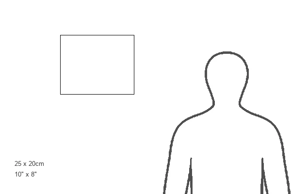
Estimated Image Size (if not cropped) is 20.3cm x 12.2cm (8" x 4.8")
Estimated Product Size is 25.4cm x 20.3cm (10" x 8")
These are individually made so all sizes are approximate
Artwork printed orientated as per the preview above, with landscape (horizontal) orientation to match the source image.
FEATURES IN THESE COLLECTIONS
> Fine Art Storehouse
> Science Inspired Art
> Illustrations & Artwork
> Fine Art Storehouse
> Science Inspired Art
> Animals
> Mammals
> Echimyidae
> Medius
> Animals
> Mammals
> Muridae
> Medius
> Animals
> Mammals
> Pteropodidae
> Medius
EDITORS COMMENTS
Capturing the intricate details of the human body, this stunning print showcases the complexity of snapping hip syndrome. The image reveals a detailed diagram of the hip joint, with its various muscles, bones, and tendons beautifully illustrated in vibrant colors against a clean white background. Snapping hip syndrome occurs when the iliopsoas tendon subluxes over the greater trochanter, causing discomfort and audible snapping or popping sensations during movement. This condition is depicted through meticulous artwork that highlights key anatomical structures such as the ilium, sacrum, lumbar vertebrae, gluteus medius muscle, and iliotibial tract. The biomedical illustration also emphasizes inflamed bursa and tendon regions associated with snapping hip syndrome. Deep trochanteric bursas and superficial trochanteric bursa are clearly labeled alongside other crucial elements like tensor fasciae latae muscle and iliopectineal eminence. With its rear view perspective allowing us to see through layers of tissue and bone structure, this print offers valuable insights into both medical professionals' understanding of this condition and individuals seeking knowledge about their own bodies. It serves as a powerful educational tool within healthcare settings or for those interested in exploring human anatomy. Lauren Shavell's artistic talent shines through her precise attention to detail in this artwork that seamlessly combines science with aesthetics.
MADE IN THE UK
Safe Shipping with 30 Day Money Back Guarantee
FREE PERSONALISATION*
We are proud to offer a range of customisation features including Personalised Captions, Color Filters and Picture Zoom Tools
SECURE PAYMENTS
We happily accept a wide range of payment options so you can pay for the things you need in the way that is most convenient for you
* Options may vary by product and licensing agreement. Zoomed Pictures can be adjusted in the Basket.