
Inflammation Collection
Inflammation: A Silent Culprit Unveiled From the excruciating pain of appendicitis to the nagging discomfort in our joints, a common yet often misunderstood phenomenon


All Professionally Made to Order for Quick Shipping
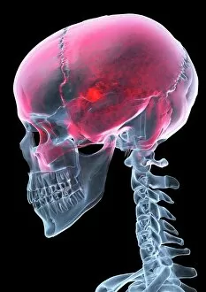
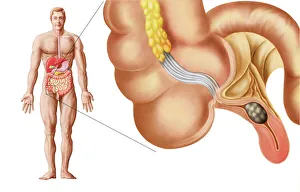
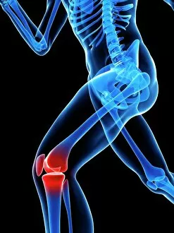
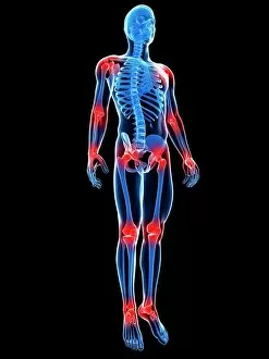
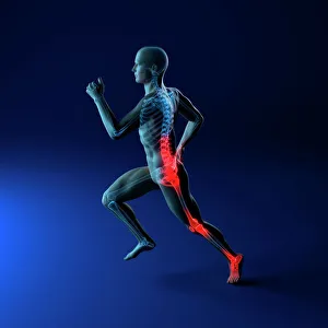
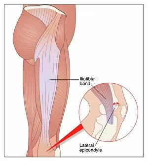
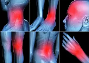
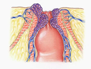
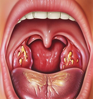
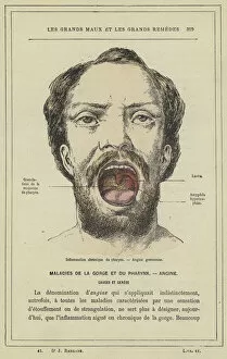
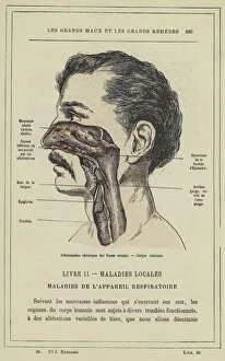
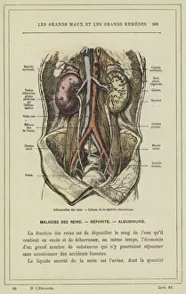
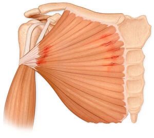
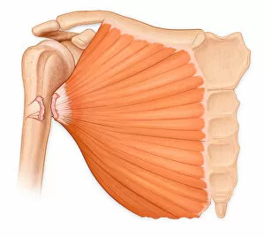
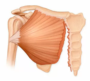
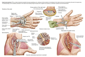
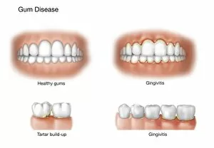
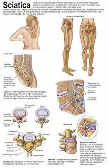
Inflammation: A Silent Culprit Unveiled From the excruciating pain of appendicitis to the nagging discomfort in our joints, a common yet often misunderstood phenomenon. This medical illustration of an inflamed appendix serves as a stark reminder of its potential dangers. Knee pain, depicted through conceptual artwork, highlights how they are hinder even the simplest movements. Whether caused by injury or conditions like arthritis, it reminds us that our knees are not invincible. Similarly, joint pain portrayed in another conceptual artwork emphasizes the debilitating effects on our mobility and quality of life. It serves as a visual representation for those who silently suffer from conditions such as rheumatoid arthritis. The intricate artwork showcasing various skin disorders draws attention to how inflammation affects not only internal organs but also external surfaces. From eczema to psoriasis, these conditions remind us that sometimes our own immune system turns against us. Headache sufferers can relate all too well to this X-ray artwork depicting their tormenting experience. Inflammation within the skull can cause intense migraines and cluster headaches that disrupt daily life and leave individuals desperate for relief. Conceptual artwork illustrating running injuries brings forth another aspect where inflammation wreaks havoc on athletes' bodies. The image captures both physical and emotional strain endured due to sprains, strains, or stress fractures caused by excessive exercise without proper recovery time. Iliotibial running injury showcased through detailed artwork demonstrates how localized they are sideline even seasoned runners. It reveals the importance of listening to one's body and seeking appropriate treatment before minor discomfort escalates into chronic agony. Body pain represented artistically symbolizes widespread inflammatory responses affecting multiple areas simultaneously. Whether due to autoimmune diseases like fibromyalgia or systemic infections, it reminds us that sometimes there is no escape from this silent culprit's grasp. Conversely, conceptual artworks portraying lower back pain highlight how specific regions may bear the brunt of inflammation.