
Biomedical Illustration Collection
"Unveiling the Intricacies of Life: Exploring Biomedical Illustration" Delving into the depths of scientific knowledge


All Professionally Made to Order for Quick Shipping
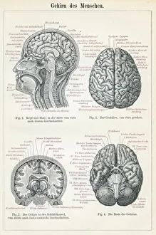
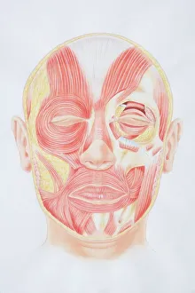
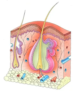
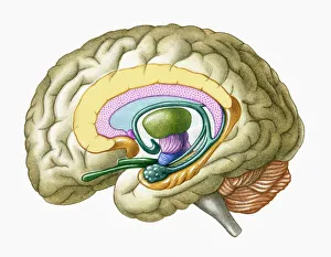
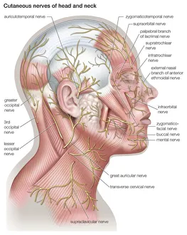
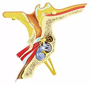
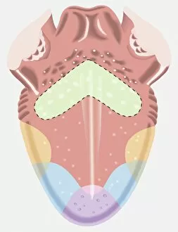
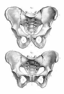
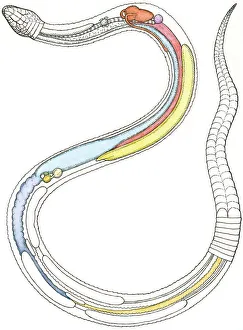
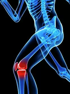
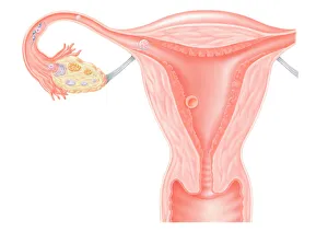
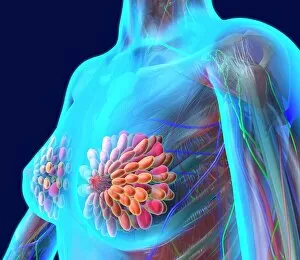
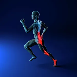
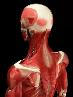
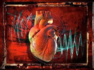
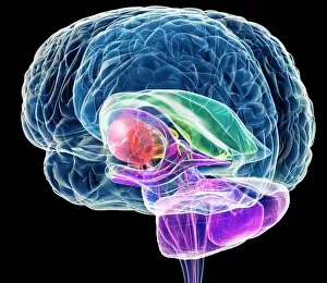
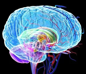
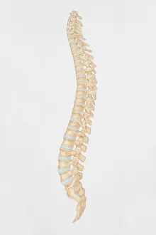
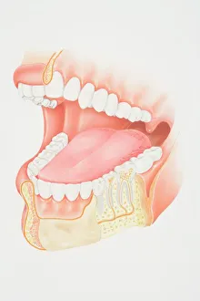
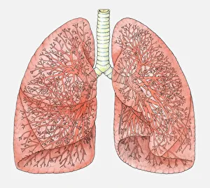
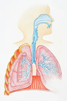
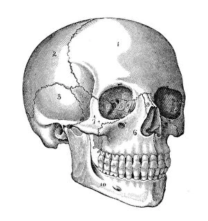
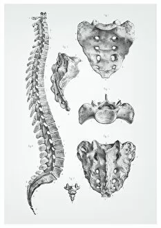
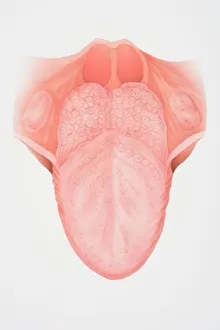
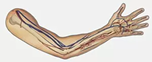
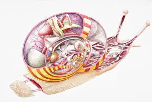
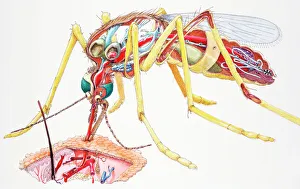
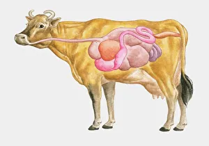
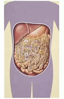
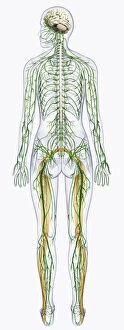
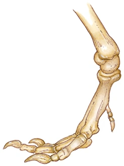
"Unveiling the Intricacies of Life: Exploring Biomedical Illustration" Delving into the depths of scientific knowledge, biomedical illustration serves as a visual gateway to unraveling the mysteries of our intricate bodies. From a brain anatomy engraving dating back to 1895, showcasing its complexity and beauty, to capturing the elegance of a DNA molecule, these illustrations encapsulate centuries worth of research and understanding. Traveling through time, we encounter an exquisite depiction from 1888 that unravels the secrets hidden within the human heart's anatomy. Moving on to a front view diagram illustrating facial muscles in meticulous detail, we witness how every twitch and expression is orchestrated by this intricate network. Venturing beneath our skin's surface, another illustration reveals the mesmerizing world of hair follicles intertwined with delicate layers of skin. Meanwhile, a cross-section illustration unveils the enigmatic limbic system and primitive forebrain within our complex cerebral landscape. Journeying further into our body's inner workings, we explore an astonishingly detailed rendering of the lymphatic system—a vital component in maintaining overall health. The male and female pelvis engraving from 1896 reminds us that even seemingly simple structures hold profound significance in shaping life itself. Stepping into auditory realms, an illustrative masterpiece showcases various components—auditory canal, eardrum, semicircular canals—intricate mechanisms responsible for sound perception. A taste map emerges next; revealing distinct regions on our tongue sensitive to bitter or sweet flavors—an artistic representation unveiling gustatory wonders. Expanding beyond human physiology lies yet another captivating realm—the internal organs of snakes—where hearts beat alongside lungs while intestines intertwine with pancreas and kidneys. This vivid portrayal highlights nature's diversity in anatomical design across species. Concluding this journey through biomedical artistry is a conceptual artwork depicting knee pain—a poignant reminder that such illustrations not only educate but also evoke empathy towards the human experience.