Fraud Blocker Skip to main content

Tendon Collection

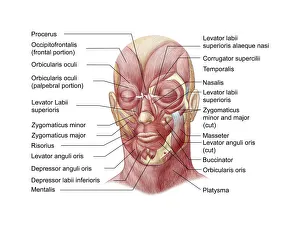
"Tendon: The Unsung Heroes of the Muscular System" The facial muscles of the human face, intricately labeled and interconnected

Background imageTendon Collection: Facial muscles of the human face (with labels)

Facial muscles of the human face (with labels)

Background imageTendon Collection: Muscular system on head

Muscular system on head
Muscular system on a head. Computer artwork showing the musculature of the side of a human males head. The muscles shown are skeletal muscles. These muscles are consciously controlled by the brain
Background imageTendon Collection: Tendinitis of the shoulder, X-ray

Tendinitis of the shoulder, X-ray
Tendinitis of the shoulder. Coloured X-ray of the shoulder of a patient showing calcification (grainy patches) around the rotator cuff tendon (upper left)

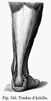
Background imageTendon Collection: Line engraving, French, 19th century

Line engraving, French, 19th century
ACHILLES TENDON. Line engraving, French, 19th century

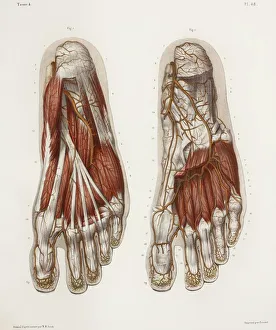
Background imageTendon Collection: Foot anatomy, 19th Century illustration

Foot anatomy, 19th Century illustration

Background imageTendon Collection: Anatomy of Organs Engraving

Anatomy of Organs Engraving
Engraved illustrations of Anatomy of the Fasciae, Integuents, and Organs of Mastication and Respiration from Iconographic Encyclopedia of Science, Literature and Art, Published in 1851

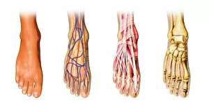
Background imageTendon Collection: Human foot anatomy showing skin, veins, arteries, muscles and bones

Human foot anatomy showing skin, veins, arteries, muscles and bones
Human foot anatomy showing skin, veins, arteries, muscles, and bones, cutaway view

Background imageTendon Collection: Hand

Hand
Antique illustration of a human hand with tendons

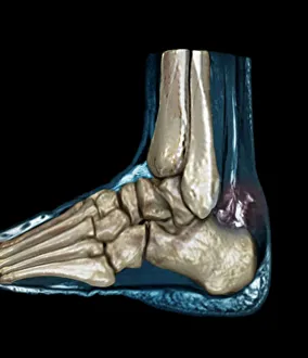
Background imageTendon Collection: Ruptured Achilles tendon, MRI C018 / 0649

Ruptured Achilles tendon, MRI C018 / 0649
Ruptured Achilles tendon. Coloured magnetic resonance imaging (MRI) scan of the foot of a 25 year old patient with a ruptured Achilles tendon

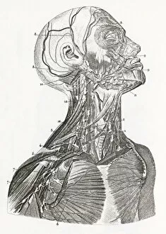
Background imageTendon Collection: Muscles of the neck

Muscles of the neck, historical artwork. The upper figure shows the front of the neck with the skin and fascia (connective tissue) removed to expose the musculature (red) and tendons

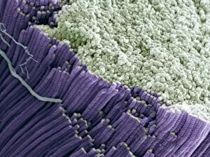
Background imageTendon Collection: Tendon, SEM

Tendon, SEM
Tendon, coloured scanning electron micrograph (SEM), showing bundles of collagen fibres. Magnification: x5000 when printed at 10 centimetres wide

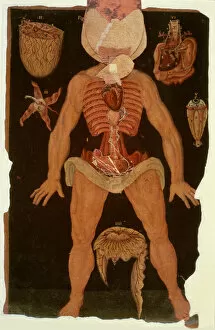
Background imageTendon Collection: The human body with superimposed colored plates by Julien Bougle

The human body with superimposed colored plates by Julien Bougle
The human body with superimposed colored plates, by Julien Bougle, circa 1899

Background imageTendon Collection: Hand anatomy, 19th Century illustration

Hand anatomy, 19th Century illustration. Historical hand coloured lithographic print showing the muscles (brown), tendons (white) and blood vessels (red and blue) of the human hand

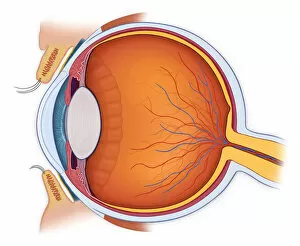
Background imageTendon Collection: Normal anatomy of the eye in cross section

Normal anatomy of the eye in cross section
Horizontal, Vision, Detail, Anatomy, Diagram, Muscle, Illustration, Lens, Iris, Medical, Eyeball, Biology, Lateral, Cut Out, White Background, Eye, Artwork, Cross Section, Color Image, Close Up

Background imageTendon Collection: Snapping hip syndrome occurs when the iliopsoas tendon subluxes over the greater

Snapping hip syndrome occurs when the iliopsoas tendon subluxes over the greater
Horizontal, Anatomy, Diagram, Muscle, Illustration, Bone, Medical, Hip, Biology, Pelvis, Femur, See Through, Rear View, Cut Out, Arthritis, White Background, Artwork, Color Image, Front View

Background imageTendon Collection: Human Brain Engraving

Human Brain Engraving
Engraved illustrations of Anatomy of the Brain and Nerves from Iconographic Encyclopedia of Science, Literature and Art, Published in 1851. Copyright has expired on this artwork. Digitally restored

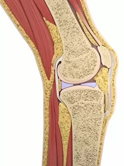
Background imageTendon Collection: Knee anatomy, artwork

Knee anatomy, artwork
Knee anatomy, computer artwork

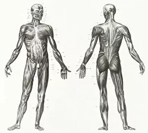
Background imageTendon Collection: Human Muscles and Ligaments Engraving

Human Muscles and Ligaments Engraving
Engraved illustrations of Anatomy of the Ligaments and Muscles from Iconographic Encyclopedia of Science, Literature and Art, Published in 1851. Copyright has expired on this artwork

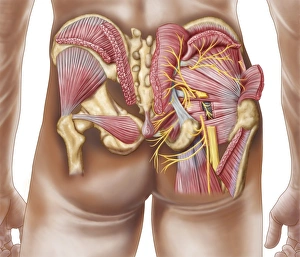
Background imageTendon Collection: Anatomy of the gluteal muscles in the human buttocks

Anatomy of the gluteal muscles in the human buttocks

Background imageTendon Collection: Muscles (engraving)

Muscles (engraving)
471461 Muscles (engraving) by Rubens, Peter Paul (1577-1640); 34.4x24 cm; Koninklijk Museum voor Schone Kunsten, Antwerp, Belgium; © Art in Flanders

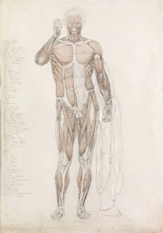
Background imageTendon Collection: Anatomical Study: Human Musculature, in Contrapposto (pencil & wash on paper)

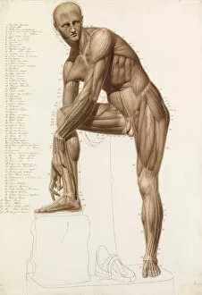
Anatomical Study: Human Musculature, in Contrapposto (pencil & wash on paper)
5889001 Anatomical Study: Human Musculature, in Contrapposto (pencil & wash on paper) by Bathe, John (1849-1874); 71.1x49.5 cm; Private Collection; (add.info)

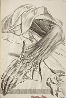
Background imageTendon Collection: Anatomia Humani Corporis by Govard Bidloo, 1685 (engraving)

Anatomia Humani Corporis by Govard Bidloo, 1685 (engraving)
2919838 Anatomia Humani Corporis by Govard Bidloo, 1685 (engraving) by Lairesse, Gerard de (1640-1711); The University of St. Andrews, Scotland, UK; (add.info.: Table 70)

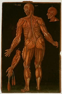
Background imageTendon Collection: Ms Hunter 364 Table X Dissection, from Anatomical Tables

Ms Hunter 364 Table X Dissection, from Anatomical Tables
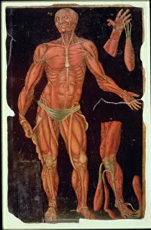
GUL174551 Ms Hunter 364 Table X Dissection, from Anatomical Tables, by John Banister (1540-1610) (oil on brown paper) by English School, (16th century); Glasgow University Library

Background imageTendon Collection: Ms Hunter 364 Table VIII Dissection, from Anatomical Tables

Ms Hunter 364 Table VIII Dissection, from Anatomical Tables
GUL174549 Ms Hunter 364 Table VIII Dissection, from Anatomical Tables, by John Banister (1540-1610) (oil on brown paper) by English School, (16th century); Glasgow University Library

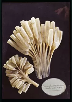
Background imageTendon Collection: Model of the muscle fibres, tendons and trachea of a fly (wax)

Model of the muscle fibres, tendons and trachea of a fly (wax)
BEN360186 Model of the muscle fibres, tendons and trachea of a fly (wax) by Susini, Clemente (1754-1814); Museo della Specola, Florence, Italy; (add.info.: Fibre muscolari)

Background imageTendon Collection: Achilles wounded by Paris, c. 1650 (oil on canvas)

Achilles wounded by Paris, c. 1650 (oil on canvas)
3930854 Achilles wounded by Paris, c.1650 (oil on canvas) by Flemalle, Bertholet (1614-75); 98x70 cm; National Museum, Stockholm

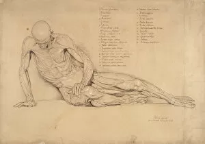
Background imageTendon Collection: An Ecorche Study of the Dying Gaul, 1840 (pen and ink on paper)

An Ecorche Study of the Dying Gaul, 1840 (pen and ink on paper)
FIT167649 An Ecorche Study of the Dying Gaul, 1840 (pen and ink on paper) by Linnell, William (1826-1906); Fitzwilliam Museum, University of Cambridge, UK; eFitzwilliam Museum; English

Background imageTendon Collection: Ms Hunter 364 Table XII Dissection (open state) from Anatomical Tables

Ms Hunter 364 Table XII Dissection (open state) from Anatomical Tables
GUL174554 Ms Hunter 364 Table XII Dissection (open state) from Anatomical Tables, by John Banister (1540-1610) (oil on brown paper) by English School, (16th century); Glasgow University Library

Background imageTendon Collection: Ms Hunter 364 Table VIII Dissection, c. 1170, from Anatomical Tables

Ms Hunter 364 Table VIII Dissection, c. 1170, from Anatomical Tables
GUL174550 Ms Hunter 364 Table VIII Dissection, c.1170, from Anatomical Tables, by John Banister (1540-1610) (oil on brown paper) by English School, (16th century); Glasgow University Library

Background imageTendon Collection: Ms Hunter 364 Table VII Dissection, from Anatomical Tables

Ms Hunter 364 Table VII Dissection, from Anatomical Tables
GUL174548 Ms Hunter 364 Table VII Dissection, from Anatomical Tables, by John Banister (1540-1610) (oil on brown paper) by English School, (16th century); Glasgow University Library

Background imageTendon Collection: Ms Hunter 364 Table XI Dissection, from Anatomical Tables

Ms Hunter 364 Table XI Dissection, from Anatomical Tables
GUL174552 Ms Hunter 364 Table XI Dissection, from Anatomical Tables, by John Banister (1540-1610) (oil on brown paper) by English School, (16th century); Glasgow University Library

Background imageTendon Collection: Anatomical Study: Human Musculature, in the Pose of Praxiteles

Anatomical Study: Human Musculature, in the Pose of Praxiteles
5889003 Anatomical Study: Human Musculature, in the Pose of Praxiteles Hermes Fastening his Sandal (pencil, pen, ink & wash on paper) by Bathe

Background imageTendon Collection: Accessory Navicular Pain Syndrome is caused by friction

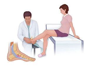
Accessory Navicular Pain Syndrome is caused by friction
Horizontal, Anatomy, Diagram, Illustration, Foot, Bone, Pressure, Injury, Medical, Doctor, Physician, Patient, Biology, Joint, Side View, See Through, Cut Out, White Background, Artwork

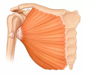
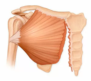
Background imageTendon Collection: Pectoralis major tear syndrome can lead to a complete rupture of the pectoralis major

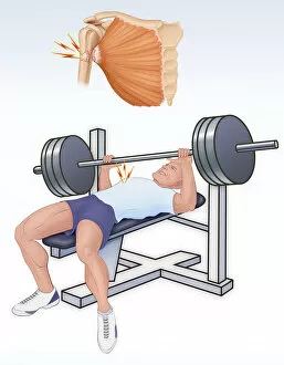
Pectoralis major tear syndrome can lead to a complete rupture of the pectoralis major
Fitness, Exercise, Strength, Anatomy, Diagram, Muscle, Illustration, Injury, Medical, Weight, Biology, Bicep, Lifting, Sportswear, Side View, Weekend Activities, Cut Out, White Background, Lifestyle

Background imageTendon Collection: Complete rupture of the pectoralis major tendon from the humerus

Complete rupture of the pectoralis major tendon from the humerus
Horizontal, Detail, Anatomy, Diagram, Muscle, Illustration, Bone, Injury, Medical, Biology, Bicep, Cut Out, White Background, Artwork, Color Image, Close Up, Healthcare And Medicine, The Human Body

Background imageTendon Collection: Os Trigonum Pain Syndrome is associated with high impact force or activites on the foot

Os Trigonum Pain Syndrome is associated with high impact force or activites on the foot
Fitness, Exercise, Anatomy, Diagram, Pain, Illustration, Bone, Injury, Medical, Biology, Sportswear, Stepping, See Through, Weekend Activities, Cut Out, White Background, Lifestyle, Vertical

Background imageTendon Collection: Pectoralis muscle tear, associated with a hematoma formation and cosmetic deformity

Pectoralis muscle tear, associated with a hematoma formation and cosmetic deformity
Horizontal, Detail, Anatomy, Diagram, Muscle, Illustration, Bone, Injury, Medical, Biology, Cut Out, White Background, Artwork, Color Image, Close Up, Healthcare And Medicine, The Human Body

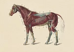
Background imageTendon Collection: External Muscles and Tendons of the Horses Body, c1879. Creator: Unknown

External Muscles and Tendons of the Horses Body, c1879. Creator: Unknown
External Muscles and Tendons of the Horses Body, c1879. From " The Book of the Horse", by S. Sidney. [Cassell & Company, Limited, London, Paris, New York, Melbourne]

Background imageTendon Collection: Cardiovascular System Engraving

Cardiovascular System Engraving
Engraved illustrations of The Vascular System from Iconographic Encyclopedia of Science, Literature and Art, Published in 1851. Copyright has expired on this artwork. Digitally restored

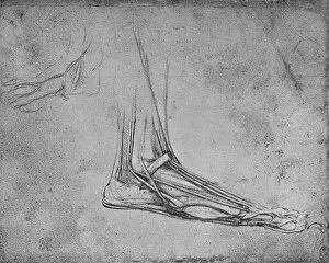
Background imageTendon Collection: Anatomical Study of Muscles of Foot, 1928. Artist: Leonardo da Vinci

Anatomical Study of Muscles of Foot, 1928. Artist: Leonardo da Vinci
Anatomical Study of Muscles of Foot, 1928. From The Mind of Leonardo Da Vinci, by Edward McCurdy. [Jonathan Cape, London, 1928]

Background imageTendon Collection: Studies of the Muscles of the Face and Arm, c1480 (1945). Artist: Leonardo da Vinci

Studies of the Muscles of the Face and Arm, c1480 (1945). Artist: Leonardo da Vinci
Studies of the Muscles of the Face and Arm, c1480 (1945). From The Drawings of Leonardo da Vinci. [Reynal & Hitchcock, New York, 1945]

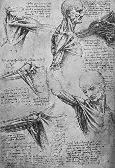
Background imageTendon Collection: Anatomical Studies of a Mans Neck and Shoulders, c1480 (1945). Artist: Leonardo da Vinci

Anatomical Studies of a Mans Neck and Shoulders, c1480 (1945). Artist: Leonardo da Vinci
Anatomical Studies of a Mans Neck and Shoulders, c1480 (1945). From The Drawings of Leonardo da Vinci. [Reynal & Hitchcock, New York, 1945]

Background imageTendon Collection: Drawings of a Left Leg Showing Bones and Tendons, c1480 (1945). Artist: Leonardo da Vinci

Drawings of a Left Leg Showing Bones and Tendons, c1480 (1945). Artist: Leonardo da Vinci
Drawings of a Left Leg Showing Bones and Tendons, c1480 (1945). From The Drawings of Leonardo da Vinci. [Reynal & Hitchcock, New York, 1945]

Background imageTendon Collection: Dissection of a Bears Foot to the Left, c1480 (1945). Artist: Leonardo da Vinci

Dissection of a Bears Foot to the Left, c1480 (1945). Artist: Leonardo da Vinci
Dissection of a Bears Foot to the Left, c1480 (1945). From The Drawings of Leonardo da Vinci. [Reynal & Hitchcock, New York, 1945]

Background imageTendon Collection: Dissection of a Bears Foot to the Right, c1480 (1945). Artist: Leonardo da Vinci

Dissection of a Bears Foot to the Right, c1480 (1945). Artist: Leonardo da Vinci
Dissection of a Bears Foot to the Right, c1480 (1945). From The Drawings of Leonardo da Vinci. [Reynal & Hitchcock, New York, 1945]

Background imageTendon Collection: Studies of Legs, of the Bones and Tendons of a Leg, of an Arm

Studies of Legs, of the Bones and Tendons of a Leg, of an Arm, and of the Bones and Tendons of an A Artist
Studies of Legs, of the Bones and Tendons of a Leg, of an Arm, and of the Bones and Tendons of an Arm and of Organs like a Valve, c1480 (1945). From The Drawings of Leonardo da Vinci

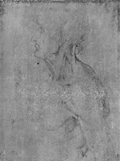
Background imageTendon Collection: Head and Bust of an Old Man to the Left, c1480 (1945). Artist: Leonardo da Vinci

Head and Bust of an Old Man to the Left, c1480 (1945). Artist: Leonardo da Vinci
Head and Bust of an Old Man to the Left, c1480 (1945). From The Drawings of Leonardo da Vinci. [Reynal & Hitchcock, New York, 1945]

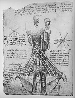
Background imageTendon Collection: Study of the Back View of a Skeleton, Showing the Tendons of the Neck, c1480 (1945)

Study of the Back View of a Skeleton, Showing the Tendons of the Neck, c1480 (1945). Artist: Leonardo da Vinci
Study of the Back View of a Skeleton, Showing the Tendons of the Neck, c1480 (1945). From The Drawings of Leonardo da Vinci. [Reynal & Hitchcock, New York, 1945]


All Professionally Made to Order for Quick Shipping

"Tendon: The Unsung Heroes of the Muscular System" The facial muscles of the human face, intricately labeled and interconnected, create a symphony of expressions that reflect our emotions. These muscles, part of the muscular system on our head, allow us to smile, frown, and convey countless other feelings. However, sometimes these they can become inflamed or irritated. Tendinitis of the shoulder is a painful condition that affects many individuals. An X-ray reveals this discomforting ailment in detail. Intriguingly captured in a line engraving from 19th-century France by an unknown artist, we witness the beauty and complexity anatomy. This illustration showcases how tendons connect muscles to bones with remarkable precision. Moving down to our feet - another masterpiece created by nature - a 19th-century illustration depicts foot anatomy in all its glory. It reveals not only skin and veins but also arteries, muscles, and bones working together harmoniously. Julien Bougle's colored plates superimposed on the human body provide an artistic representation showcasing various systems within us. Among them are tendons that play their crucial role silently yet efficiently. Delving deeper into internal structures brings us to an exquisite engraving depicting the intricate anatomy of organs. Here we see how tendons intertwine with other vital components like blood vessels and nerves to ensure proper functioning throughout our bodies. Zooming back out again to examine foot anatomy once more - this time showing skin texture alongside veins, arteries, muscles, and bones - we gain further appreciation for these unsung heroes called tendons. Our hands too rely heavily on healthy tendons for dexterity and strength as they perform daily tasks ranging from delicate movements to powerful grips. They enable us to write words like these or hold loved ones close with tender care. Yet even strong they can be vulnerable; an MRI image captures a ruptured Achilles tendon revealing just how crucial their integrity is for our mobility and overall well-being.