
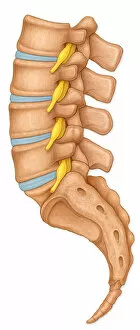
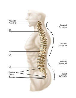
Lumbar Vertebrae Collection
The lumbar vertebrae, located in the lower back region of the spine, play a crucial role in providing stability and support to our body


All Professionally Made to Order for Quick Shipping
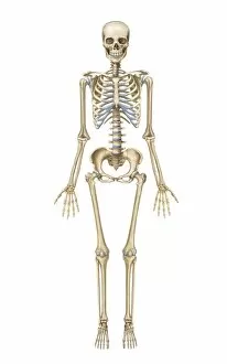
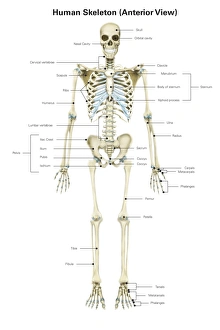
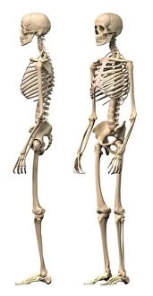
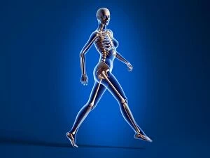
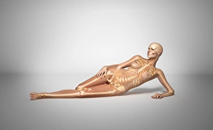
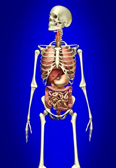
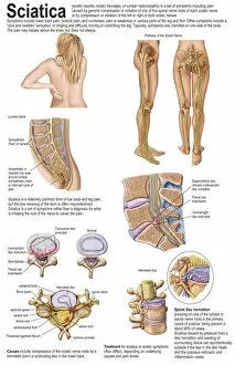
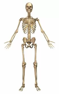
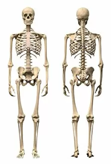
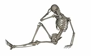
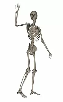
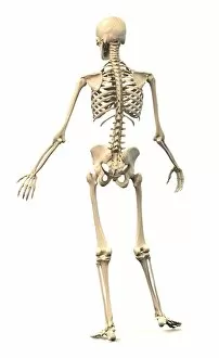
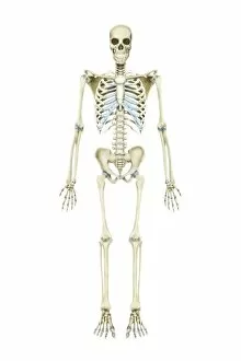
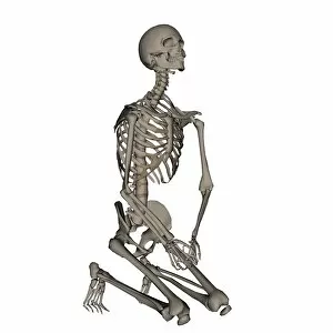
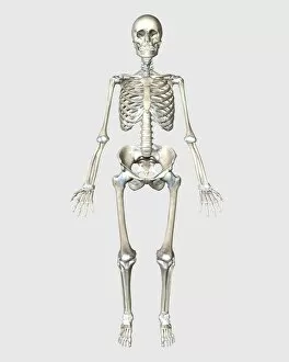
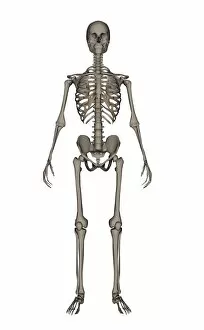
The lumbar vertebrae, located in the lower back region of the spine, play a crucial role in providing stability and support to our body. In a normal lateral view, these vertebrae showcase the intricate network of spinal nerve roots that branch out from them. However, repetitive activities like aerobics that involve lifting up the knee can potentially lead to trauma in this area. One condition associated with such activities is snapping hip syndrome, where the iliopsoas tendon subluxes over the greater trochanter causing discomfort and pain. This highlights how important it is to be mindful of our movements during exercise routines. When we visualize the human skeletal system from different angles - front view or anterior view - we gain a deeper understanding of its complexity. The male anatomy provides us with side views and perspective views of their skeleton, allowing us to appreciate its structure even more. Examining a posterior view reveals not only the hip bones but also showcases the gluteus maximus muscle along with lumbar involvement. It reminds us that muscles work hand-in-hand with bones for optimal functioning. To truly grasp how everything fits together within our bodies, superimposed images help immensely. Whether it's a female body with bone skeleton and internal organs overlaid or a naked woman lying down showcasing her skeletal framework, these visuals provide an insightful look into our inner workings. Taking another approach by focusing solely on specific areas like the vertebral column gives us an opportunity to explore left lateral views exclusively dedicated to understanding this vital part of our anatomy. Lastly, an anterior view specifically highlighting labels allows for easy identification and comprehension when studying human pelvis structures while incorporating both male skeletons against blue backgrounds alongside internal organs adds depth to anatomical knowledge. Exploring various aspects related to lumbar vertebrae broadens our awareness about their significance in maintaining bodily functions while emphasizing caution during physical activities that may cause harm if not performed correctly.